Generic placeholder image

Letters in Drug Design & Discovery

Editor-in-Chief

ISSN (Print): 1570-1808
ISSN (Online): 1875-628X

Research Article

Exploring Energy Profiles of Protein-Protein Interactions (PPIs) Using DFT Method

Author(s): Sanket Bapat, Renu Vyas* and Muthukumarasamy Karthikeyan

Volume 16, Issue 6, 2019

Page: [670 - 677] Pages: 8

DOI: 10.2174/1570180815666180815151141

Price: $65

Abstract

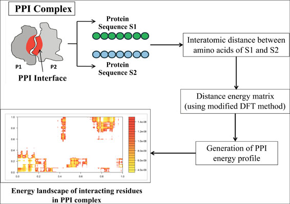
Background: Large-scale energy landscape characterization of protein-protein interactions (PPIs) is important to understand the interaction mechanism and protein-protein docking methods. The experimental methods for detecting energy landscapes are tedious and the existing computational methods require longer simulation time.

Objective: The objective of the present work is to ascertain the energy profiles at the interface regions in a rapid manner to analyze the energy landscape of protein-protein interactions.

Methods: The atomic coordinates obtained from the X-ray and NMR spectroscopy data are considered as inputs to compute cumulative energy profiles for experimentally validated protein-protein complexes. The energies computed by the program were comparable to the standard molecular dynamics simulations.

Results: The PPI Profiler not only enables rapid generation of energy profiles but also facilitates the detection of hot spot residue atoms involved therein.

Conclusion: The hotspot residues and their computed energies matched with the experimentally determined hot spot residues and their energies which correlated well by employing the MM/GBSA method. The proposed method can be employed to scan entire proteomes across species at an atomic level to study the key PPI interactions.

Keywords: Protein-protein interaction, energy profiles, DFT, energy landscape, hot spot residues, proteomes.

Graphical Abstract
[1]
Kshirsagar, M.; Murugesan, K.; Carbonell, J.G.; Klein-Seetharaman, J. Multitask matrix completion for learning protein interactions across diseases. J. Comput. Biol., 2017, 24(6), 501-514.
[2]
Tsai, M.Y.; Zheng, W.; Balamurugan, D.; Schafer, N.P.; Kim, B.L.; Cheung, M.S.; Wolynes, P.G. Electrostatics, structure prediction, and the energy landscapes for protein folding and binding. Protein Sci., 2016, 25(1), 255-269.
[3]
Conway, P.; Tyka, M.D.; DiMaio, F.; Konerding, D.E.; Baker, D. Relaxation of backbone bond geometry improves protein energy landscape modeling. Protein Sci., 2014, 23(1), 47-55.
[4]
Moritsugu, K.; Terada, T.; Kidera, A. Energy landscape of allatom protein-protein interactions revealed by multiscale enhanced sampling. PLoS Comput. Biol, 2014, 10(10), e1003901/1-e100390.
[5]
Ikebe, J.; Sakuraba, S.; Kono, H. Adaptive lambda square dynamics simulation: An efficient conformational sampling method for biomolecules. J. Comput. Chem., 2014, 35(1), 39-50.
[6]
Pfaendtner, J.; Bonomi, M. Efficient sampling of high-dimensional free-energy landscapes with parallel bias metadynamics. J. Chem. Theory Comput., 2015, 11(11), 5062-5067.
[7]
Pfaendtner, J.; Bonomi, M. Efficient sampling of high-dimensional free-energy landscapes with parallel bias metadynamics. J. Chem. Theory Comput., 2015, 11(11), 5062-5067.
[8]
Barducci, A.; Bonomi, M.; Prakash, M.K.; Parrinello, M. Free-energy landscape of protein oligomerization from atomistic simulations. Proc. Natl. Acad. Sci. USA, 2013, 110, 49.
[9]
Zheng, F.; Jewell, H.; Fitzpatrick, J.; Zhang, J.; Mierke, D.F.; Grigoryan, G. Computational design of selective peptides to discriminate between similar PDZ domains in an oncogenic pathway. J. Mol. Biol., 2015, 427(2), 491-510.
[10]
Plattner, N.; Doerr, S.; De Fabritiis, G.; Noé, F. Complete protein-protein association kinetics in atomic detail revealed by molecular dynamics simulations and Markov modelling. Nat. Chem., 2015, 9(10), e1005.
[11]
Saba-El-Leil, M.K.; Frémin, C.; Meloche, S. Redundancy in the world of MAP kinases: All for one. Front. Cell Dev. Biol., 2016, 4, 67.
[12]
Ribone, S.R.; Quevedo, M.A. Structural basis for the potent inhibition of the HIV integrase-LEDGF/p75 protein-protein interaction. J. Mol. Graph. Model, 2017, 189-198, DOI: 10.1016/j.jmgm.2017. 05.019.
[13]
Guo, W.; Wisniewski, J.A.; Ji, H. Hot spot-based design of small-molecule inhibitors for protein-protein interactions. ACS Med. Chem. Lett., 2014, 24(11), 2546-2554.
[14]
Radoux, C.J.; Olsson, T.S.; Pitt, W.R.; Groom, C.R.; Blundell, T.L. Identifying interactions that determine fragment binding at protein hotspots. J. Med. Chem., 2016, 59(9), 4314-4325.
[15]
Zhao, P.; Qin, K.; Ye, X.; Wang, Y.; and Chen, Y. A trajectory clustering approach based on decision graph and data field for detecting hotspots. Int. J. Geogr. Inf. Sci., 2017, 31(6), 1101-1127.
[16]
Glowacki, D.R.; Rodgers, W.J.; Shannon, R.; Robertson, S.H.; Harvey, J.N. Reaction and relaxation at surface hotspots: Using molecular dynamics and the energy-grained master equation to describe diamond etching. Phil. Trans. R. Soc. A., 2017, 375(2092), 20160206.
[17]
Vyas, R.; Bapat, S.; Goel, P.; Karthikeyan, M.; Tambe, S.S.; Kulkarni, B.D. Application of Genetic Programming (GP) formalism for building disease predictive models from protein-protein interactions (PPI) data. IEEE/ACM Trans. Comput. Biol. Bioinformatics, 2016, 15(1), 27-37.
[http://dx.doi.org/10.1109/TCBB.2016.2621042]
[18]
Rakers, C.; Bermudez, M.; Keller, B.G.; Mortier, J.; Wolber, G. Computational close up on protein-protein interactions: How to unravel the invisible using molecular dynamics simulations? Wiley Interdiscip. Rev. Comput. Mol. Sci., 2015, 5(5), 345-359.
[19]
Karthikeyan, M.; Vyas, R. A method for classifying protein interactions using energy profile fingerprinting. 2015, 2015-NF-0168. (Sourceforge link: https://sourceforge.net/projects/ppienergy/? source =directory).
[20]
Grimme, S. Density functional theory with London dispersion corrections. Wiley Interdiscip. Rev. Comput. Mol. Sci., 2011, 1(2), 211-228.
[21]
Jones, S.; Thornton, J.M. Principles of protein-protein interactions. Proc. Natl. Acad. Sci. USA, 1996, 93(1), 13-20.
[22]
Pelay‐Gimeno, M.; Glas, A.; Koch, O.; Grossmann, T.N. Structure‐based design of inhibitors of protein-protein interactions: Mimicking peptide binding epitopes. Angew. Chem. Int. Ed. Engl., 2015, 54(31), 8896-8927.
[23]
Yang, X.; Coulombe-Huntington, J.; Kang, S.; Sheynkman, G.M.; Hao, T.; Richardson, A.; Sun, S.; Yang, F.; Shen, Y.A.; Murray, R.R.; Spirohn, K. Widespread expansion of protein interaction capabilities by alternative splicing. Cell, 2016, 164(4), 805-817.
[24]
Giabbai, B.; Degano, M. Crystal structure to 1.7 Å of the Escherichia coli pyrimidine nucleoside hydrolase YeiK, a novel candidate for cancer gene therapy. Structure, 2004, 12(5), 739-749.
[25]
Hikita, H.; Takehara, T. Regulation of apoptosis by Bcl-2 family proteins in liver injury. Molecules Systems and Signaling in Liver Injury, 2017, (75-85). Springer, Cham.
[26]
O’Connell, K.; Jinks-Robertson, S.; Petes, T.D. Elevated genome-wide instability in yeast mutants lacking RNase H activity. Genetics, 2015, 201(3), 963-975.
[27]
Carter-Su, C.; Schwartz, J.; Argetsinger, L.S. Growth hormone signaling pathways. Growth Hormone I.G.F. Res., 2016, 28, 11-15.
[28]
Balamurugan, K. HIF‐1 at the crossroads of hypoxia, inflammation, and cancer. Int. J. Cancer, 2016, 138(5), 1058-1066.
[29]
Perilla, J.R.; Goh, B.C.; Cassidy, C.K.; Liu, B.; Bernardi, R.C.; Rudack, T.; Yu, H.; Wu, Z.; Schulten, K. Molecular dynamics simulations of large macromolecular complexes. Curr. Opin. Struct. Biol., 2015, 31, 64-74.
[30]
Tóth, F.; Kádas, J.; Mótyán, J.A.; Tőzsér, J. Effect of internal cleavage site mutations in human immunodeficiency virus type 1 capsid protein on its structure and function. FEBS Open Bio, 2016, 6(8), 847-859.
[31]
Biertümpfel, C.; Yang, W.; Suck, D. Crystal structure of T4 endonuclease VII resolving a Holliday junction. Nature, 2007, 449(7162), 616.
[32]
Harvey, A.L. Cardiotoxins from cobra venoms. Handbook Nat. Toxins, 2018, 85-106.
[33]
Fujinaga, M.; Sielecki, A.R.; Read, R.J.; Ardelt, W.; Laskowski, M.; James, M.N.G. Crystal and molecular structures of the complex of alphachymotrypsin with its inhibitor turkey ovomucoid third domain at 1.8 Amstrong resolution. J. Mol. Biol., 1987, 195(2), 397-418.
[34]
Song, H.K.; Suh, S.W. Kunitz-type soybean trypsin inhibitor revisited: Refined structure of its complex with porcine trypsin reveals an insight into the interaction between a homologous inhibitor from Erythrinacaffra and tissue-type plasminogen activator. J. Mol. Biol., 1998, 275(2), 347-363.
[35]
Fischmann, T.O.; Bentley, G.A.; Bhat, T.N.; Boulot, G.; Mariuzza, R.A.; Phillips, S.E.; Tello, D.; Poljak, R.J. Crystallographic refinement of the three-dimensional structure of the FabD1. 3-lysozyme complex at 2.5-A resolution. J. Biochem., 1991, 266(20), 12915-12920.
[36]
Mena-Ulecia, K.; Tiznado, W.; Caballero, J. Study of the differential activity of thrombin inhibitors using docking, QSAR, molecular dynamics, and MM-GBSA. PLoS One, 2015, 10(11), e0142774.
[37]
Genheden, S.; Ryde, U. The MM/PBSA and MM/GBSA methods to estimate ligand-binding affinities. Expert Opin. Drug Discov., 2015, 10(5), 449-461.
[38]
Van der Spoel, D.; Lindahl, E.; Hess, B. The GROMACS development team, GROMACS user manual, version 4.6. 5 , 2013.
[39]
Thorn, K.S.; Bogan, A.A. ASEdb: A database of alanine mutations and their effects on the free energy of binding in protein interactions. Bioinformatics, 2001, 17(3), 284-285.

Rights & Permissions Print Cite
© 2024 Bentham Science Publishers | Privacy Policy