Review Article

Gene Therapy in the Anterior Eye Segment

Author(s): Cynthia Amador, Ruchi Shah, Sean Ghiam, Andrei A. Kramerov and Alexander V. Ljubimov*

Volume 22, Issue 2, 2022

Published on: 23 April, 2021

Page: [104 - 131] Pages: 28

DOI: 10.2174/1566523221666210423084233

open access plus

Abstract

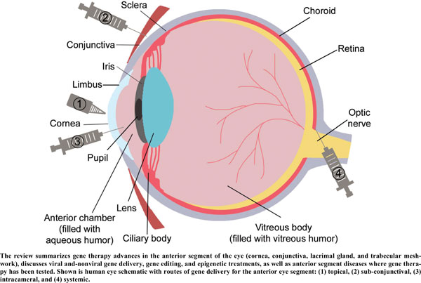
This review provides comprehensive information about the advances in gene therapy in the anterior segment of the eye, including cornea, conjunctiva, lacrimal gland, and trabecular meshwork. We discuss gene delivery systems, including viral and non-viral vectors as well as gene editing techniques, mainly CRISPR-Cas9, and epigenetic treatments, including antisense and siRNA therapeutics. We also provide a detailed analysis of various anterior segment diseases where gene therapy has been tested with corresponding outcomes. Disease conditions include corneal and conjunctival fibrosis and scarring, corneal epithelial wound healing, corneal graft survival, corneal neovascularization, genetic corneal dystrophies, herpetic keratitis, glaucoma, dry eye disease, and other ocular surface diseases. Although most of the analyzed results on the use and validity of gene therapy at the ocular surface have been obtained in vitro or using animal models, we also discuss the available human studies. Gene therapy approaches are currently considered very promising as emerging future treatments of various diseases, and this field is rapidly expanding.

Keywords: Gene therapy, cornea, corneal dystrophy, corneal wound healing, keratitis, corneal neovascularization, glaucoma, dry eye, graft survival, non-viral vector, nanoconstruct, drug delivery, adenovirus, adeno-associated virus, retrovirus, lentivirus, antisense, siRNA, CRISPR-Cas9.

Next »
Graphical Abstract
[1]
Anguela XM, High KA. Entering the modern era of gene therapy. Annu Rev Med 2019; 70: 273-88.
[http://dx.doi.org/10.1146/annurev-med-012017-043332] [PMID: 30477394]
[2]
Wirth T, Parker N, Ylä-Herttuala S. History of gene therapy. Gene 2013; 525(2): 162-9.
[http://dx.doi.org/10.1016/j.gene.2013.03.137] [PMID: 23618815]
[3]
Dunbar CE, High KA, Joung JK, Kohn DB, Ozawa K, Sadelain M. Gene therapy comes of age. Science 2018; 359(6372): eaan4672.
[http://dx.doi.org/10.1126/science.aan4672] [PMID: 29326244]
[4]
High KA, Roncarolo MG. Gene Therapy. N Engl J Med 2019; 381(5): 455-64.
[http://dx.doi.org/10.1056/NEJMra1706910] [PMID: 31365802]
[5]
Bennett J, Maguire AM. Gene therapy for ocular disease. Mol Ther 2000; 1(6): 501-5.
[http://dx.doi.org/10.1006/mthe.2000.0080] [PMID: 10933974]
[6]
Borrás T. Recent developments in ocular gene therapy. Exp Eye Res 2003; 76(6): 643-52.
[http://dx.doi.org/10.1016/S0014-4835(03)00030-7] [PMID: 12742346]
[7]
Di Iorio E, Barbaro V, Alvisi G, et al. New frontiers of corneal gene therapy. Hum Gene Ther 2019; 30(8): 923-45.
[http://dx.doi.org/10.1089/hum.2019.026] [PMID: 31020856]
[8]
Ljubimov AV. Overview of gene therapy in anterior segment. Invest Ophthalmol Vis Sci 2019; 60: 1037.
[9]
Mohan RR, Martin LM, Sinha NR. Novel insights into gene therapy in the cornea. Exp Eye Res 2021; 202: 108361.
[http://dx.doi.org/10.1016/j.exer.2020.108361] [PMID: 33212142]
[10]
Shirley JL, de Jong YP, Terhorst C, Herzog RW. Immune responses to viral gene therapy vectors. Mol Ther 2020; 28(3): 709-22.
[http://dx.doi.org/10.1016/j.ymthe.2020.01.001] [PMID: 31968213]
[11]
Volpers C, Kochanek S. Adenoviral vectors for gene transfer and therapy. J Gene Med 2004; 6(Suppl. 1): S164-71.
[http://dx.doi.org/10.1002/jgm.496] [PMID: 14978759]
[12]
Mohan RR, Rodier JT, Sharma A. Corneal gene therapy: Basic science and translational perspective. Ocul Surf 2013; 11(3): 150-64.
[http://dx.doi.org/10.1016/j.jtos.2012.10.004] [PMID: 23838017]
[13]
Saghizadeh M, Kramerov AA, Yu FS, Castro MG, Ljubimov AV. Normalization of wound healing and diabetic markers in organ cultured human diabetic corneas by adenoviral delivery of c-Met gene. Invest Ophthalmol Vis Sci 2010; 51(4): 1970-80.
[http://dx.doi.org/10.1167/iovs.09-4569] [PMID: 19933191]
[14]
Lai CM, Brankov M, Zaknich T, et al. Inhibition of angiogenesis by adenovirus-mediated sFlt-1 expression in a rat model of corneal neovascularization. Hum Gene Ther 2001; 12(10): 1299-310.
[http://dx.doi.org/10.1089/104303401750270959] [PMID: 11440623]
[15]
Kaufmann C, Mortimer LA, Brereton HM, et al. Interleukin-10 gene transfer in rat limbal transplantation. Curr Eye Res 2017; 42(11): 1426-34.
[http://dx.doi.org/10.1080/02713683.2017.1344714] [PMID: 28925732]
[16]
Bessis N, GarciaCozar FJ, Boissier MC. Immune responses to gene therapy vectors: Influence on vector function and effector mechanisms. Gene Ther 2004; 11(Suppl. 1): S10-7.
[http://dx.doi.org/10.1038/sj.gt.3302364] [PMID: 15454952]
[17]
Kochanek S. High-capacity adenoviral vectors for gene transfer and somatic gene therapy. Hum Gene Ther 1999; 10(15): 2451-9.
[http://dx.doi.org/10.1089/10430349950016807] [PMID: 10543611]
[18]
Boutin S, Monteilhet V, Veron P, et al. Prevalence of serum IgG and neutralizing factors against adeno-associated virus (AAV) types 1, 2, 5, 6, 8, and 9 in the healthy population: Implications for gene therapy using AAV vectors. Hum Gene Ther 2010; 21(6): 704-12.
[http://dx.doi.org/10.1089/hum.2009.182] [PMID: 20095819]
[19]
Hareendran S, Balakrishnan B, Sen D, Kumar S, Srivastava A, Jayandharan GR. Adeno-associated virus (AAV) vectors in gene therapy: Immune challenges and strategies to circumvent them. Rev Med Virol 2013; 23(6): 399-413.
[http://dx.doi.org/10.1002/rmv.1762] [PMID: 24023004]
[20]
Mingozzi F, High KA. Immune responses to AAV vectors: Overcoming barriers to successful gene therapy. Blood 2013; 122(1): 23-36.
[http://dx.doi.org/10.1182/blood-2013-01-306647] [PMID: 23596044]
[21]
Martino AT, Markusic DM. Immune response mechanisms against AAV vectors in animal models. Mol Ther Methods Clin Dev 2019; 17: 198-208.
[http://dx.doi.org/10.1016/j.omtm.2019.12.008] [PMID: 31970198]
[22]
Naso MF, Tomkowicz B, Perry WL III, Strohl WR. Adeno-associated virus (AAV) as a vector for gene therapy. BioDrugs 2017; 31(4): 317-34.
[http://dx.doi.org/10.1007/s40259-017-0234-5] [PMID: 28669112]
[23]
Mohan RR, Tovey JC, Sharma A, Schultz GS, Cowden JW, Tandon A. Targeted decorin gene therapy delivered with adeno-associated virus effectively retards corneal neovascularization in vivo. PLoS One 2011; 6(10): e26432.
[http://dx.doi.org/10.1371/journal.pone.0026432] [PMID: 22039486]
[24]
Sharma A, Tovey JC, Ghosh A, Mohan RR. AAV serotype influences gene transfer in corneal stroma in vivo. Exp Eye Res 2010; 91(3): 440-8.
[http://dx.doi.org/10.1016/j.exer.2010.06.020] [PMID: 20599959]
[25]
Liu J, Saghizadeh M, Tuli SS, et al. Different tropism of adenoviruses and adeno-associated viruses to corneal cells: Implications for corneal gene therapy. Mol Vis 2008; 14: 2087-96.
[PMID: 19023450]
[26]
Song L, Llanga T, Conatser LM, Zaric V, Gilger BC, Hirsch ML. Serotype survey of AAV gene delivery via subconjunctival injection in mice. Gene Ther 2018; 25(6): 402-14.
[http://dx.doi.org/10.1038/s41434-018-0035-6] [PMID: 30072815]
[27]
Bastola P, Song L, Gilger BC, Hirsch ML. Adeno-associated virus mediated gene therapy for corneal diseases. Pharmaceutics 2020; 12(8): 767.
[http://dx.doi.org/10.3390/pharmaceutics12080767] [PMID: 32823625]
[28]
Tarallo V, Bogdanovich S, Hirano Y, et al. Inhibition of choroidal and corneal pathologic neovascularization by Plgf1-de gene transfer. Invest Ophthalmol Vis Sci 2012; 53(13): 7989-96.
[http://dx.doi.org/10.1167/iovs.12-10658] [PMID: 23139276]
[29]
Saint-Geniez M, Maharaj AS, Walshe TE, et al. Endogenous VEGF is required for visual function: Evidence for a survival role on müller cells and photoreceptors. PLoS One 2008; 3(11): e3554.
[http://dx.doi.org/10.1371/journal.pone.0003554] [PMID: 18978936]
[30]
Lu Y, Tai PWL, Ai J, et al. Transcriptome profiling of neovascularized corneas reveals miR-204 as a multi-target biotherapy deliverable by rAAVs. Mol Ther Nucleic Acids 2018; 10: 349-60.
[http://dx.doi.org/10.1016/j.omtn.2017.12.019] [PMID: 29499946]
[31]
Hirsch ML, Conatser LM, Smith SM, et al. AAV vector-meditated expression of HLA-G reduces injury-induced corneal vascularization, immune cell infiltration, and fibrosis. Sci Rep 2017; 7(1): 17840.
[http://dx.doi.org/10.1038/s41598-017-18002-9] [PMID: 29259248]
[32]
Parker DG, Brereton HM, Coster DJ, Williams KA. The potential of viral vector-mediated gene transfer to prolong corneal allograft survival. Curr Gene Ther 2009; 9(1): 33-44.
[http://dx.doi.org/10.2174/156652309787354621] [PMID: 19275570]
[33]
Vance M, Llanga T, Bennett W, et al. AAV gene therapy for MPS1-associated corneal blindness. Sci Rep 2016; 6: 22131.
[http://dx.doi.org/10.1038/srep22131] [PMID: 26899286]
[34]
Gupta S, Rodier JT, Sharma A, et al. Targeted AAV5-Smad7 gene therapy inhibits corneal scarring in vivo. PLoS One 2017; 12(3): e0172928.
[http://dx.doi.org/10.1371/journal.pone.0172928] [PMID: 28339457]
[35]
Zhou L, Zhu X, Tan J, Wang J, Xing Y. Effect of recombinant adeno-associated virus mediated transforming growth factor-β1 on corneal allograft survival after high-risk penetrating keratoplasty. Transpl Immunol 2013; 28(4): 164-9.
[http://dx.doi.org/10.1016/j.trim.2013.04.007] [PMID: 23624044]
[36]
Alvarez-Rivera F, Rey-Rico A, Venkatesan JK, et al. Controlled release of rAAV vectors from APMA-functionalized contact lenses for corneal gene therapy. Pharmaceutics 2020; 12(4): 335.
[http://dx.doi.org/10.3390/pharmaceutics12040335] [PMID: 32283694]
[37]
Cockrell AS, Kafri T. Gene delivery by lentivirus vectors. Mol Biotechnol 2007; 36(3): 184-204.
[http://dx.doi.org/10.1007/s12033-007-0010-8] [PMID: 17873406]
[38]
Sakuma T, Barry MA, Ikeda Y. Lentiviral vectors: Basic to translational. Biochem J 2012; 443(3): 603-18.
[http://dx.doi.org/10.1042/BJ20120146] [PMID: 22507128]
[39]
Parker M, Bellec J, McFarland T, et al. Suppression of neovascularization of donor corneas by transduction with equine infectious anemia virus-based lentiviral vectors expressing endostatin and angiostatin. Hum Gene Ther 2014; 25(5): 408-18.
[http://dx.doi.org/10.1089/hum.2013.079] [PMID: 24460027]
[40]
Wang T, Zhou XT, Yu Y, et al. Inhibition of corneal fibrosis by Smad7 in rats after photorefractive keratectomy. Chin Med J (Engl) 2013; 126(8): 1445-50.
[PMID: 23595375]
[41]
Oliveira LA, Kim C, Sousa LB, Schwab IR, Rosenblatt MI. Gene transfer to primary corneal epithelial cells with an integrating lentiviral vector. Arq Bras Oftalmol 2010; 73(5): 447-53.
[http://dx.doi.org/10.1590/S0004-27492010000500012] [PMID: 21225131]
[42]
Ramamoorth M, Narvekar A. Non viral vectors in gene therapy- an overview. J Clin Diagn Res 2015; 9(1): GE01-6.
[http://dx.doi.org/10.7860/JCDR/2015/10443.5394] [PMID: 25738007]
[43]
Hardee CL, Arévalo-Soliz LM, Hornstein BD, Zechiedrich L. Advances in non-viral DNA vectors for gene therapy. Genes (Basel) 2017; 8(2): 65.
[http://dx.doi.org/10.3390/genes8020065] [PMID: 28208635]
[44]
Kampik D, Ali RR, Larkin DF. Experimental gene transfer to the corneal endothelium. Exp Eye Res 2012; 95(1): 54-9.
[http://dx.doi.org/10.1016/j.exer.2011.07.001] [PMID: 21777585]
[45]
Reyes-Sandoval A, Ertl HC. CpG methylation of a plasmid vector results in extended transgene product expression by circumventing induction of immune responses. Mol Ther 2004; 9(2): 249-61.
[http://dx.doi.org/10.1016/j.ymthe.2003.11.008] [PMID: 14759809]
[46]
Krieg AM, Wu T, Weeratna R, et al. Sequence motifs in adenoviral DNA block immune activation by stimulatory CpG motifs. Proc Natl Acad Sci USA 1998; 95(21): 12631-6.
[http://dx.doi.org/10.1073/pnas.95.21.12631] [PMID: 9770537]
[47]
Yew NS, Zhao H, Wu IH, et al. Reduced inflammatory response to plasmid DNA vectors by elimination and inhibition of immunostimulatory CpG motifs. Mol Ther 2000; 1(3): 255-62.
[http://dx.doi.org/10.1006/mthe.2000.0036] [PMID: 10933941]
[48]
de Wolf HK, Johansson N, Thong AT, et al. Plasmid CpG depletion improves degree and duration of tumor gene expression after intravenous administration of polyplexes. Pharm Res 2008; 25(7): 1654-62.
[http://dx.doi.org/10.1007/s11095-008-9558-7] [PMID: 18317886]
[49]
Lee H, Park K. In vitro cytotoxicity of zinc oxide nanoparticles in cultured Statens Serum institut rabbit cornea cells. Toxicol Res 2019; 35(3): 287-94.
[http://dx.doi.org/10.5487/TR.2019.35.3.287] [PMID: 31341558]
[50]
Tandon A, Sharma A, Rodier JT, Klibanov AM, Rieger FG, Mohan RR. BMP7 gene transfer via gold nanoparticles into stroma inhibits corneal fibrosis in vivo. PLoS One 2013; 8(6): e66434.
[http://dx.doi.org/10.1371/journal.pone.0066434] [PMID: 23799103]
[51]
Masse F, Desjardins P, Ouellette M, et al. Synthesis of ultrastable gold nanoparticles as a new drug delivery system. Molecules 2019; 24(16): 2929.
[http://dx.doi.org/10.3390/molecules24162929] [PMID: 31412609]
[52]
Tartaj P, Morales M, Veintemillas-Verdaguer S, González-Carreño T, Serna CJ. The preparation of magnetic nanoparticles for applications in biomedicine. J Phys D Appl Phys 2003; 36: R182-97.
[http://dx.doi.org/10.1088/0022-3727/36/13/202]
[53]
Cornell LE, Wehmeyer JL, Johnson AJ, Desilva MN, Zamora DO. Magnetic nanoparticles as a potential vehicle for corneal endothelium repair. Mil Med 2016; 181(5)(Suppl.): 232-9.
[http://dx.doi.org/10.7205/MILMED-D-15-00151] [PMID: 27168578]
[54]
Xia X, Atkins M, Dalal R, et al. Magnetic human corneal endothelial cell transplant: Delivery, retention, and short-term efficacy. Invest Ophthalmol Vis Sci 2019; 60(7): 2438-48.
[http://dx.doi.org/10.1167/iovs.18-26001] [PMID: 31158276]
[55]
Moysidis SN, Alvarez-Delfin K, Peschansky VJ, et al. Magnetic field-guided cell delivery with nanoparticle-loaded human corneal endothelial cells. Nanomedicine (Lond) 2015; 11(3): 499-509.
[http://dx.doi.org/10.1016/j.nano.2014.12.002] [PMID: 25596075]
[56]
Tong YC, Chang SF, Liu CY, Kao WW, Huang CH, Liaw J. Eye drop delivery of nano-polymeric micelle formulated genes with cornea-specific promoters. J Gene Med 2007; 9(11): 956-66.
[http://dx.doi.org/10.1002/jgm.1093] [PMID: 17724775]
[57]
Cholkar K, Hariharan S, Gunda S, Mitra AK. Optimization of dexamethasone mixed nanomicellar formulation. AAPS PharmSciTech 2014; 15(6): 1454-67.
[http://dx.doi.org/10.1208/s12249-014-0159-y] [PMID: 24980081]
[58]
Luis de Redín I, Boiero C, Recalde S, et al. In vivo effect of bevacizumab-loaded albumin nanoparticles in the treatment of corneal neovascularization. Exp Eye Res 2019; 185: 107697.
[http://dx.doi.org/10.1016/j.exer.2019.107697] [PMID: 31228461]
[59]
Jiang M, Gan L, Zhu C, Dong Y, Liu J, Gan Y. Cationic core-shell liponanoparticles for ocular gene delivery. Biomaterials 2012; 33(30): 7621-30.
[http://dx.doi.org/10.1016/j.biomaterials.2012.06.079] [PMID: 22789720]
[60]
Ljubimova JY, Sun T, Mashouf L, et al. Covalent nano delivery systems for selective imaging and treatment of brain tumors. Adv Drug Deliv Rev 2017; 113: 177-200.
[http://dx.doi.org/10.1016/j.addr.2017.06.002] [PMID: 28606739]
[61]
Kopeček J, Yang J. Polymer nanomedicines. Adv Drug Deliv Rev 2020; 156: 40-64.
[http://dx.doi.org/10.1016/j.addr.2020.07.020] [PMID: 32735811]
[62]
Tong YC, Chang SF, Kao WW, Liu CY, Liaw J. Polymeric micelle gene delivery of bcl-xL via eye drop reduced corneal apoptosis following epithelial debridement. J Control Release 2010; 147(1): 76-83.
[http://dx.doi.org/10.1016/j.jconrel.2010.06.006] [PMID: 20600407]
[63]
Kalomiraki M, Thermos K, Chaniotakis NA. Dendrimers as tunable vectors of drug delivery systems and biomedical and ocular applications. Int J Nanomedicine 2015; 11: 1-12.
[PMID: 26730187]
[64]
Holden CA, Tyagi P, Thakur A, et al. Polyamidoamine dendrimer hydrogel for enhanced delivery of antiglaucoma drugs. Nanomedicine (Lond) 2012; 8(5): 776-83.
[http://dx.doi.org/10.1016/j.nano.2011.08.018] [PMID: 21930109]
[65]
Soiberman U, Kambhampati SP, Wu T, et al. Subconjunctival injectable dendrimer-dexamethasone gel for the treatment of corneal inflammation. Biomaterials 2017; 125: 38-53.
[http://dx.doi.org/10.1016/j.biomaterials.2017.02.016] [PMID: 28226245]
[66]
Yang X, Wang L, Li L, et al. A novel dendrimer-based complex co-modified with cyclic RGD hexapeptide and penetratin for noninvasive targeting and penetration of the ocular posterior segment. Drug Deliv 2019; 26(1): 989-1001.
[http://dx.doi.org/10.1080/10717544.2019.1667455] [PMID: 31571502]
[67]
Maloy SR, Hughes KT. Microinjection.Brenner’s encyclopedia of genetics. Amsterdam: Elsevier/Academic Press 2013.
[68]
Kim B, Lee S, Suvas S, Rouse BT. Application of plasmid DNA encoding IL-18 diminishes development of herpetic stromal keratitis by antiangiogenic effects. J Immunol 2005; 175(1): 509-16.
[http://dx.doi.org/10.4049/jimmunol.175.1.509] [PMID: 15972686]
[69]
Jani PD, Singh N, Jenkins C, et al. Nanoparticles sustain expression of Flt intraceptors in the cornea and inhibit injury-induced corneal angiogenesis. Invest Ophthalmol Vis Sci 2007; 48(5): 2030-6.
[http://dx.doi.org/10.1167/iovs.06-0853] [PMID: 17460257]
[70]
Galiacy SD, Fournié P, Massoudi D, et al. Matrix metalloproteinase 14 overexpression reduces corneal scarring. Gene Ther 2011; 18(5): 462-8.
[http://dx.doi.org/10.1038/gt.2010.159] [PMID: 21160532]
[71]
He Z, Pipparelli A, Manissolle C, et al. Ex vivo gene electrotransfer to the endothelium of organ cultured human corneas. Ophthalmic Res 2010; 43(1): 43-55.
[http://dx.doi.org/10.1159/000246577] [PMID: 19829011]
[72]
Blair-Parks K, Weston BC, Dean DA. High-level gene transfer to the cornea using electroporation. J Gene Med 2002; 4(1): 92-100.
[http://dx.doi.org/10.1002/jgm.231] [PMID: 11828392]
[73]
Rosazza C, Meglic SH, Zumbusch A, Rols MP, Miklavcic D. Gene electrotransfer: A mechanistic perspective. Curr Gene Ther 2016; 16(2): 98-129.
[http://dx.doi.org/10.2174/1566523216666160331130040] [PMID: 27029943]
[74]
Yu WZ, Li XX, She HC, et al. Gene transfer of kringle 5 of plasminogen by electroporation inhibits corneal neovascularization. Ophthalmic Res 2003; 35(5): 239-46.
[http://dx.doi.org/10.1159/000072143] [PMID: 12920335]
[75]
Wells DJ. Gene therapy progress and prospects: Electroporation and other physical methods. Gene Ther 2004; 11(18): 1363-9.
[http://dx.doi.org/10.1038/sj.gt.3302337] [PMID: 15295618]
[76]
Liu S, Romano V, Steger B, Kaye SB, Hamill KJ, Willoughby CE. Gene-based antiangiogenic applications for corneal neovascularization. Surv Ophthalmol 2018; 63(2): 193-213.
[http://dx.doi.org/10.1016/j.survophthal.2017.10.006] [PMID: 29080632]
[77]
Berdugo M, Valamanesh F, Andrieu C, et al. Delivery of antisense oligonucleotide to the cornea by iontophoresis. Antisense Nucleic Acid Drug Dev 2003; 13(2): 107-14.
[http://dx.doi.org/10.1089/108729003321629647] [PMID: 12804037]
[78]
Sekijima H, Ehara J, Hanabata Y, et al. Characterization of ocular iontophoretic drug transport of ionic and non-ionic compounds in isolated rabbit cornea and conjunctiva. Biol Pharm Bull 2016; 39(6): 959-68.
[http://dx.doi.org/10.1248/bpb.b15-00932] [PMID: 27040754]
[79]
Vinciguerra P, Romano V, Rosetta P, et al. Iontophoresis-assisted corneal collagen cross-linking with epithelial debridement: Preliminary results. BioMed Res Int 2016; 2016: 3720517.
[http://dx.doi.org/10.1155/2016/3720517] [PMID: 27547758]
[80]
Bouheraoua N, Jouve L, Borderie V, Laroche L. Three different protocols of corneal collagen crosslinking in keratoconus: Conventional, accelerated and iontophoresis. J Vis Exp 2015; 105(105): 53119.
[http://dx.doi.org/10.3791/53119] [PMID: 26650390]
[81]
Bikbova G, Bikbov M. Transepithelial corneal collagen cross-linking by iontophoresis of riboflavin. Acta Ophthalmol 2014; 92(1): e30-4.
[http://dx.doi.org/10.1111/aos.12235] [PMID: 23848196]
[82]
Zhang EP, Muller A, Schulte F, et al. Minimizing side effects of ballistic gene transfer into the murine corneal epithelium. Graefes Arch Clin Exp Ophthalmol 2002; 240(2): 114-9.
[http://dx.doi.org/10.1007/s00417-001-0411-5] [PMID: 11931076]
[83]
Zagon IS, Sassani JW, Verderame MF, McLaughlin PJ. Particle-mediated gene transfer of opioid growth factor receptor cDNA regulates cell proliferation of the corneal epithelium. Cornea 2005; 24(5): 614-9.
[http://dx.doi.org/10.1097/01.ico.0000153561.89902.57] [PMID: 15968171]
[84]
Zagon IS, Sassani JW, Malefyt KJ, McLaughlin PJ. Regulation of corneal repair by particle-mediated gene transfer of opioid growth factor receptor complementary DNA. Arch Ophthalmol 2006; 124(11): 1620-4.
[http://dx.doi.org/10.1001/archopht.124.11.1620] [PMID: 17102011]
[85]
König Merediz SA, Zhang EP, Wittig B, Hoffmann F. Ballistic transfer of minimalistic immunologically defined expression constructs for IL4 and CTLA4 into the corneal epithelium in mice after orthotopic corneal allograft transplantation. Graefes Arch Clin Exp Ophthalmol 2000; 238(8): 701-7.
[http://dx.doi.org/10.1007/s004170000144] [PMID: 11011692]
[86]
Müller A, Zhang EP, Schroff M, Wittig B, Hoffmann F. Influence of ballistic gene transfer on antigen-presenting cells in murine corneas. Graefes Arch Clin Exp Ophthalmol 2002; 240(10): 851-9.
[http://dx.doi.org/10.1007/s00417-002-0536-1] [PMID: 12397435]
[87]
Mohan RR, Sharma A, Netto MV, Sinha S, Wilson SE. Gene therapy in the cornea. Prog Retin Eye Res 2005; 24(5): 537-59.
[http://dx.doi.org/10.1016/j.preteyeres.2005.04.001] [PMID: 15955719]
[88]
Mohan RR, Tovey JC, Sharma A, Tandon A. Gene therapy in the cornea: 2005--present. Prog Retin Eye Res 2012; 31(1): 43-64.
[http://dx.doi.org/10.1016/j.preteyeres.2011.09.001] [PMID: 21967960]
[89]
Bemelmans AP, Arsenijevic Y, Majo F. Efficient lentiviral gene transfer into corneal stroma cells using a femtosecond laser. Gene Ther 2009; 16(7): 933-8.
[http://dx.doi.org/10.1038/gt.2009.41] [PMID: 19387484]
[90]
Jumelle C, Mauclair C, Houzet J, et al. Delivery of macromolecules into the endothelium of whole ex vivo human cornea by femtosecond laser-activated carbon nanoparticles. Br J Ophthalmol 2016; 100(8): 1151-6.
[http://dx.doi.org/10.1136/bjophthalmol-2015-307610] [PMID: 27226345]
[91]
Toropainen E, Hornof M, Kaarniranta K, Johansson P, Urtti A. Corneal epithelium as a platform for secretion of transgene products after transfection with liposomal gene eyedrops. J Gene Med 2007; 9(3): 208-16.
[http://dx.doi.org/10.1002/jgm.1011] [PMID: 17351984]
[92]
Dannowski H, Bednarz J, Reszka R, Engelmann K, Pleyer U. Lipid-mediated gene transfer of acidic fibroblast growth factor into human corneal endothelial cells. Exp Eye Res 2005; 80(1): 93-101.
[http://dx.doi.org/10.1016/j.exer.2004.08.024] [PMID: 15652530]
[93]
Rossi A, Kontarakis Z, Gerri C, et al. Genetic compensation induced by deleterious mutations but not gene knockdowns. Nature 2015; 524(7564): 230-3.
[http://dx.doi.org/10.1038/nature14580] [PMID: 26168398]
[94]
Scoles DR, Minikel EV, Pulst SM. Antisense oligonucleotides: A primer. Neurol Genet 2019; 5(2): e323.
[http://dx.doi.org/10.1212/NXG.0000000000000323] [PMID: 31119194]
[95]
Cursiefen C, Viaud E, Bock F, et al. Aganirsen antisense oligonucleotide eye drops inhibit keratitis-induced corneal neovascularization and reduce need for transplantation: The I-CAN study. Ophthalmology 2014; 121(9): 1683-92.
[http://dx.doi.org/10.1016/j.ophtha.2014.03.038] [PMID: 24811963]
[96]
Cursiefen C, Bock F, Horn FK, et al. GS-101 antisense oligonucleotide eye drops inhibit corneal neovascularization: Interim results of a randomized phase II trial. Ophthalmology 2009; 116(9): 1630-7.
[http://dx.doi.org/10.1016/j.ophtha.2009.04.016] [PMID: 19643487]
[97]
Wasmuth S, Bauer D, Yang Y, Steuhl KP, Heiligenhaus A. Topical treatment with antisense oligonucleotides targeting tumor necrosis factor-alpha in herpetic stromal keratitis. Invest Ophthalmol Vis Sci 2003; 44(12): 5228-34.
[http://dx.doi.org/10.1167/iovs.03-0312] [PMID: 14638721]
[98]
Elbadawy HM, Gailledrat M, Desseaux C, Ponzin D, Ferrari S. Targeting herpetic keratitis by gene therapy. J Ophthalmol 2012; 2012: 594869.
[http://dx.doi.org/10.1155/2012/594869] [PMID: 23326647]
[99]
Hu J, Rong Z, Gong X, et al. Oligonucleotides targeting TCf4 triplet repeat expansion inhibit RNA foci and mis-splicing in Fuchs’ dystrophy. Hum Mol Genet 2018; 27(6): 1015-26.
[http://dx.doi.org/10.1093/hmg/ddy018] [PMID: 29325021]
[100]
Kramerov AA, Shah R, Ding H, et al. Novel nanopolymer RNA therapeutics normalize human diabetic corneal wound healing and epithelial stem cells. Nanomedicine (Lond) 2020; 32: 102332.
[http://dx.doi.org/10.1016/j.nano.2020.102332] [PMID: 33181273]
[101]
Zarouchlioti C, Sanchez-Pintado B, Hafford Tear NJ, et al. Antisense therapy for a common corneal dystrophy ameliorates TCf4 repeat expansion-mediated toxicity. Am J Hum Genet 2018; 102(4): 528-39.
[http://dx.doi.org/10.1016/j.ajhg.2018.02.010] [PMID: 29526280]
[102]
Gibson DJ, Tuli SS, Schultz GS. Dual-phase iontophoresis for the delivery of antisense oligonucleotides. Nucleic Acid Ther 2017; 27(4): 238-50.
[http://dx.doi.org/10.1089/nat.2016.0654] [PMID: 28375679]
[103]
Chau VQ, Hu J, Gong X, et al. Delivery of antisense oligonucleotides to the cornea. Nucleic Acid Ther 2020; 30(4): 207-14.
[http://dx.doi.org/10.1089/nat.2019.0838] [PMID: 32202944]
[104]
Supe S, Upadhya A, Singh K. Role of small interfering RNA (siRNA) in targeting ocular neovascularization: A review. Exp Eye Res 2021; 202: 108329.
[http://dx.doi.org/10.1016/j.exer.2020.108329] [PMID: 33198953]
[105]
Liu Q, Wu K, Qiu X, Yang Y, Lin X, Yu M. siRNA silencing of gene expression in trabecular meshwork: RhoA siRNA reduces IOP in mice. Curr Mol Med 2012; 12(8): 1015-27.
[http://dx.doi.org/10.2174/156652412802480907] [PMID: 22741561]
[106]
Courtney DG, Atkinson SD, Allen EH, et al. siRNA silencing of the mutant keratin 12 allele in corneal limbal epithelial cells grown from patients with Meesmann’s epithelial corneal dystrophy. Invest Ophthalmol Vis Sci 2014; 55(5): 3352-60.
[http://dx.doi.org/10.1167/iovs.13-12957] [PMID: 24801514]
[107]
Qin Q, Shi Y, Zhao Q, et al. Effects of CD25siRNA gene transfer on high-risk rat corneal graft rejection. Graefes Arch Clin Exp Ophthalmol 2015; 253(10): 1765-76.
[http://dx.doi.org/10.1007/s00417-015-3067-2] [PMID: 26024991]
[108]
Guzman-Aranguez A, Loma P, Pintor J. Small-interfering RNAs (siRNAs) as a promising tool for ocular therapy. Br J Pharmacol 2013; 170(4): 730-47.
[http://dx.doi.org/10.1111/bph.12330] [PMID: 23937539]
[109]
Saghizadeh M, Epifantseva I, Hemmati DM, Ghiam CA, Brunken WJ, Ljubimov AV. Enhanced wound healing, kinase and stem cell marker expression in diabetic organ-cultured human corneas upon MMP-10 and cathepsin F gene silencing. Invest Ophthalmol Vis Sci 2013; 54(13): 8172-80.
[http://dx.doi.org/10.1167/iovs.13-13233] [PMID: 24255036]
[110]
Saghizadeh M, Dib CM, Brunken WJ, Ljubimov AV. Normalization of wound healing and stem cell marker patterns in organ-cultured human diabetic corneas by gene therapy of limbal cells. Exp Eye Res 2014; 129: 66-73.
[http://dx.doi.org/10.1016/j.exer.2014.10.022] [PMID: 25446319]
[111]
Schiroli D, Gómara MJ, Maurizi E, et al. Effective in vivo topical delivery of siRNA and gene silencing in intact corneal epithelium using a modified cell-penetrating peptide. Mol Ther Nucleic Acids 2019; 17: 891-906.
[http://dx.doi.org/10.1016/j.omtn.2019.07.017] [PMID: 31476668]
[112]
Baran-Rachwalska P, Torabi-Pour N, Sutera FM, et al. Topical siRNA delivery to the cornea and anterior eye by hybrid silicon-lipid nanoparticles. J Control Release 2020; 326: 192-202.
[http://dx.doi.org/10.1016/j.jconrel.2020.07.004] [PMID: 32653503]
[113]
Yu W, Wu Z. Ocular delivery of CRISPR/Cas genome editing components for treatment of eye diseases. Adv Drug Deliv Rev 2021; 168: 181-95.
[http://dx.doi.org/10.1016/j.addr.2020.06.011] [PMID: 32603815]
[114]
Wu Z, Yang H, Colosi P. Effect of genome size on AAV vector packaging. Mol Ther 2010; 18(1): 80-6.
[http://dx.doi.org/10.1038/mt.2009.255] [PMID: 19904234]
[115]
Suzuki T, Sasaki T, Yano K, et al. Development of a recombinant adenovirus vector production system free of replication-competent adenovirus by utilizing a packaging size limit of the viral genome. Virus Res 2011; 158(1-2): 154-60.
[http://dx.doi.org/10.1016/j.virusres.2011.03.026] [PMID: 21470569]
[116]
Balaggan KS, Ali RR. Ocular gene delivery using lentiviral vectors. Gene Ther 2012; 19(2): 145-53.
[http://dx.doi.org/10.1038/gt.2011.153] [PMID: 22052240]
[117]
Yang Y, Wang L, Bell P, et al. A dual AAV system enables the Cas9-mediated correction of a metabolic liver disease in newborn mice. Nat Biotechnol 2016; 34(3): 334-8.
[http://dx.doi.org/10.1038/nbt.3469] [PMID: 26829317]
[118]
Jain A, Zode G, Kasetti RB, et al. CRISPR-Cas9-based treatment of myocilin-associated glaucoma. Proc Natl Acad Sci USA 2017; 114(42): 11199-204.
[http://dx.doi.org/10.1073/pnas.1706193114] [PMID: 28973933]
[119]
Holmgaard A, Askou AL, Benckendorff JNE, et al. In vivo knockout of the VEGFA gene by lentiviral delivery of CRISPR/Cas9 in mouse retinal pigment epithelium cells. Mol Ther Nucleic Acids 2017; 9: 89-99.
[http://dx.doi.org/10.1016/j.omtn.2017.08.016] [PMID: 29246327]
[120]
Lai Y, Yue Y, Duan D. Evidence for the failure of adeno-associated virus serotype 5 to package a viral genome > or = 8.2 kb. Mol Ther 2010; 18(1): 75-9.
[http://dx.doi.org/10.1038/mt.2009.256] [PMID: 19904238]
[121]
Wang S, Sengel C, Emerson MM, Cepko CL. A gene regulatory network controls the binary fate decision of rod and bipolar cells in the vertebrate retina. Dev Cell 2014; 30(5): 513-27.
[http://dx.doi.org/10.1016/j.devcel.2014.07.018] [PMID: 25155555]
[122]
Bakondi B, Lv W, Lu B, et al. In vivo CRISPR/Cas9 gene editing corrects retinal dystrophy in the S334ter-3 rat model of autosomal dominant retinitis pigmentosa. Mol Ther 2016; 24(3): 556-63.
[http://dx.doi.org/10.1038/mt.2015.220] [PMID: 26666451]
[123]
Chen G, Abdeen AA, Wang Y, et al. A biodegradable nanocapsule delivers a Cas9 ribonucleoprotein complex for in vivo genome editing. Nat Nanotechnol 2019; 14(10): 974-80.
[http://dx.doi.org/10.1038/s41565-019-0539-2] [PMID: 31501532]
[124]
Wang Y, Shahi PK, Xie R, et al. A pH-responsive silica-metal-organic framework hybrid nanoparticle for the delivery of hydrophilic drugs, nucleic acids, and CRISPR-Cas9 genome-editing machineries. J Control Release 2020; 324: 194-203.
[http://dx.doi.org/10.1016/j.jconrel.2020.04.052] [PMID: 32380204]
[125]
Gaudana R, Ananthula HK, Parenky A, Mitra AK. Ocular drug delivery. AAPS J 2010; 12(3): 348-60.
[http://dx.doi.org/10.1208/s12248-010-9183-3] [PMID: 20437123]
[126]
Yellepeddi VK, Palakurthi S. Recent Advances in Topical Ocular Drug Delivery. J Ocul Pharmacol Ther 2016; 32(2): 67-82.
[http://dx.doi.org/10.1089/jop.2015.0047] [PMID: 26666398]
[127]
Joseph RR, Venkatraman SS. Drug delivery to the eye: What benefits do nanocarriers offer? Nanomedicine (Lond) 2017; 12(6): 683-702.
[http://dx.doi.org/10.2217/nnm-2016-0379] [PMID: 28186436]
[128]
Gote V, Sikder S, Sicotte J, Pal D. Ocular drug delivery: Present innovations and future challenges. J Pharmacol Exp Ther 2019; 370(3): 602-24.
[http://dx.doi.org/10.1124/jpet.119.256933] [PMID: 31072813]
[129]
Rafiei F, Tabesh H, Farzad F. Sustained subconjunctival drug delivery systems: Current trends and future perspectives. Int Ophthalmol 2020; 40(9): 2385-401.
[http://dx.doi.org/10.1007/s10792-020-01391-8] [PMID: 32383131]
[130]
Barar J, Javadzadeh AR, Omidi Y. Ocular novel drug delivery: Impacts of membranes and barriers. Expert Opin Drug Deliv 2008; 5(5): 567-81.
[http://dx.doi.org/10.1517/17425247.5.5.567] [PMID: 18491982]
[131]
Bourne RRA, Flaxman SR, Braithwaite T, et al. Magnitude, temporal trends, and projections of the global prevalence of blindness and distance and near vision impairment: A systematic review and meta-analysis. Lancet Glob Health 2017; 5(9): e888-97.
[http://dx.doi.org/10.1016/S2214-109X(17)30293-0] [PMID: 28779882]
[132]
World Health Organization Report. Visual impairment and blindness fact sheet. 2019. Available from: https://www.who.int/news-room/fact-sheets/detail/blindness-and-visual-impairment
[133]
Shu DY, Lovicu FJ. Myofibroblast transdifferentiation: The dark force in ocular wound healing and fibrosis. Prog Retin Eye Res 2017; 60: 44-65.
[http://dx.doi.org/10.1016/j.preteyeres.2017.08.001] [PMID: 28807717]
[134]
Myofibroblasts HB. Exp Eye Res 2016; 142: 56-70.
[http://dx.doi.org/10.1016/j.exer.2015.07.009] [PMID: 26192991]
[135]
Mohan RR, Gupta R, Mehan MK, Cowden JW, Sinha S. Decorin transfection suppresses profibrogenic genes and myofibroblast formation in human corneal fibroblasts. Exp Eye Res 2010; 91(2): 238-45.
[http://dx.doi.org/10.1016/j.exer.2010.05.013] [PMID: 20546727]
[136]
Schaefer L, Iozzo RV. Biological functions of the small leucine-rich proteoglycans: From genetics to signal transduction. J Biol Chem 2008; 283(31): 21305-9.
[http://dx.doi.org/10.1074/jbc.R800020200] [PMID: 18463092]
[137]
Yamaguchi Y, Mann DM, Ruoslahti E. Negative regulation of transforming growth factor-beta by the proteoglycan decorin. Nature 1990; 346(6281): 281-4.
[http://dx.doi.org/10.1038/346281a0] [PMID: 2374594]
[138]
Harper JR, Spiro RC, Gaarde WA, et al. Role of transforming growth factor beta and decorin in controlling fibrosis. Methods Enzymol 1994; 245: 241-54.
[http://dx.doi.org/10.1016/0076-6879(94)45014-5] [PMID: 7760736]
[139]
Huijun W, Long C, Zhigang Z, Feng J, Muyi G. Ex vivo transfer of the decorin gene into rat glomerulus via a mesangial cell vector suppressed extracellular matrix accumulation in experimental glomerulonephritis. Exp Mol Pathol 2005; 78(1): 17-24.
[http://dx.doi.org/10.1016/j.yexmp.2004.07.006] [PMID: 15596056]
[140]
Donnelly KS, Giuliano EA, Sharma A, Tandon A, Rodier JT, Mohan RR. Decorin-PEI nanoconstruct attenuates equine corneal fibroblast differentiation. Vet Ophthalmol 2014; 17(3): 162-9.
[http://dx.doi.org/10.1111/vop.12060] [PMID: 23718145]
[141]
Sharma A, Rodier JT, Tandon A, Klibanov AM, Mohan RR. Attenuation of corneal myofibroblast development through nanoparticle-mediated soluble transforming growth factor-β type II receptor (sTGFβRII) gene transfer. Mol Vis 2012; 18: 2598-607.
[PMID: 23112572]
[142]
Wang S, Hirschberg R. BMP7 antagonizes TGF-β -dependent fibrogenesis in mesangial cells. Am J Physiol Renal Physiol 2003; 284(5): f1006-13.
[http://dx.doi.org/10.1152/ajprenal.00382.2002] [PMID: 12676736]
[143]
Wang S, Hirschberg R. Bone morphogenetic protein-7 signals opposing transforming growth factor beta in mesangial cells. J Biol Chem 2004; 279(22): 23200-6.
[http://dx.doi.org/10.1074/jbc.M311998200] [PMID: 15047707]
[144]
Nakao A, Afrakhte M, Morén A, et al. Identification of Smad7, a TGFbeta-inducible antagonist of TGF-β signalling. Nature 1997; 389(6651): 631-5.
[http://dx.doi.org/10.1038/39369] [PMID: 9335507]
[145]
Saika S, Ikeda K, Yamanaka O, et al. Therapeutic effects of adenoviral gene transfer of bone morphogenic protein-7 on a corneal alkali injury model in mice. Lab Invest 2005; 85(4): 474-86.
[http://dx.doi.org/10.1038/labinvest.3700247] [PMID: 15696184]
[146]
Gupta S, Fink MK, Ghosh A, et al. Novel combination BMP7 and HGF gene therapy instigates selective myofibroblast apoptosis and reduces corneal haze in vivo. Invest Ophthalmol Vis Sci 2018; 59(2): 1045-57.
[http://dx.doi.org/10.1167/iovs.17-23308] [PMID: 29490341]
[147]
Cordeiro MF, Reichel MB, Gay JA, D’Esposita F, Alexander RA, Khaw PT. Transforming growth factor-β1, -β2, and -β3 in vivo: Effects on normal and mitomycin C-modulated conjunctival scarring. Invest Ophthalmol Vis Sci 1999; 40(9): 1975-82.
[PMID: 10440251]
[148]
Cordeiro MF, Mead A, Ali RR, et al. Novel antisense oligonucleotides targeting TGF-beta inhibit in vivo scarring and improve surgical outcome. Gene Ther 2003; 10(1): 59-71.
[http://dx.doi.org/10.1038/sj.gt.3301865] [PMID: 12525838]
[149]
Chun YY, Yap ZL, Seet LF, et al. Positive-charge tuned gelatin hydrogel-siSPARC injectable for siRNA anti-scarring therapy in post glaucoma filtration surgery. Sci Rep 2021; 11(1): 1470.
[http://dx.doi.org/10.1038/s41598-020-80542-4] [PMID: 33446775]
[150]
Saika S, Yamanaka O, Okada Y, et al. Effect of overexpression of PPARgamma on the healing process of corneal alkali burn in mice. Am J Physiol Cell Physiol 2007; 293(1): C75-86.
[http://dx.doi.org/10.1152/ajpcell.00332.2006] [PMID: 17625041]
[151]
Behrens A, Gordon EM, Li L, et al. Retroviral gene therapy vectors for prevention of excimer laser-induced corneal haze. Invest Ophthalmol Vis Sci 2002; 43(4): 968-77.
[PMID: 11923236]
[152]
Ljubimov AV, Saghizadeh M. Progress in corneal wound healing. Prog Retin Eye Res 2015; 49: 17-45.
[http://dx.doi.org/10.1016/j.preteyeres.2015.07.002] [PMID: 26197361]
[153]
Saghizadeh M, Brown DJ, Castellon R, et al. Overexpression of matrix metalloproteinase-10 and matrix metalloproteinase-3 in human diabetic corneas: A possible mechanism of basement membrane and integrin alterations. Am J Pathol 2001; 158(2): 723-34.
[http://dx.doi.org/10.1016/S0002-9440(10)64015-1] [PMID: 11159210]
[154]
Saghizadeh M, Kramerov AA, Tajbakhsh J, et al. Proteinase and growth factor alterations revealed by gene microarray analysis of human diabetic corneas. Invest Ophthalmol Vis Sci 2005; 46(10): 3604-15.
[http://dx.doi.org/10.1167/iovs.04-1507] [PMID: 16186340]
[155]
Daniels JT, Limb GA, Saarialho-Kere U, Murphy G, Khaw PT. Human corneal epithelial cells require MMP-1 for HGF-mediated migration on collagen I. Invest Ophthalmol Vis Sci 2003; 44(3): 1048-55.
[http://dx.doi.org/10.1167/iovs.02-0442] [PMID: 12601028]
[156]
Bevan D, Gherardi E, Fan TP, Edwards D, Warn R. Diverse and potent activities of HGF/SF in skin wound repair. J Pathol 2004; 203(3): 831-8.
[http://dx.doi.org/10.1002/path.1578] [PMID: 15221943]
[157]
Kakazu A, Chandrasekher G, Bazan HE. HGF protects corneal epithelial cells from apoptosis by the PI-3K/Akt-1/Bad- but not the ERK1/2-mediated signaling pathway. Invest Ophthalmol Vis Sci 2004; 45(10): 3485-92.
[http://dx.doi.org/10.1167/iovs.04-0372] [PMID: 15452053]
[158]
Neuss S, Becher E, Wöltje M, Tietze L, Jahnen-Dechent W. Functional expression of HGF and HGF receptor/c-met in adult human mesenchymal stem cells suggests a role in cell mobilization, tissue repair, and wound healing. Stem Cells 2004; 22(3): 405-14.
[http://dx.doi.org/10.1634/stemcells.22-3-405] [PMID: 15153617]
[159]
Chmielowiec J, Borowiak M, Morkel M, et al. c-Met is essential for wound healing in the skin. J Cell Biol 2007; 177(1): 151-62.
[http://dx.doi.org/10.1083/jcb.200701086] [PMID: 17403932]
[160]
Saghizadeh M, Kramerov AA, Yaghoobzadeh Y, et al. Adenovirus-driven overexpression of proteinases in organ-cultured normal human corneas leads to diabetic-like changes. Brain Res Bull 2010; 81(2-3): 262-72.
[http://dx.doi.org/10.1016/j.brainresbull.2009.10.007] [PMID: 19828126]
[161]
Kramerov AA, Saghizadeh M, Ljubimov AV. Adenoviral gene therapy for diabetic keratopathy: Effects on wound healing and stem cell marker expression in human organ-cultured corneas and limbal epithelial cells. J Vis Exp 2016; 110(110): e54058.
[http://dx.doi.org/10.3791/54058] [PMID: 27077448]
[162]
Sassani JW, Mc Laughlin PJ, Zagon IS. The Yin and Yang of the opioid growth regulatory system: Focus on diabetes-The Lorenz E. Zimmerman Tribute Lecture. J Diabetes Res 2016; 2016: 9703729.
[http://dx.doi.org/10.1155/2016/9703729] [PMID: 27703986]
[163]
Funari VA, Winkler M, Brown J, Dimitrijevich SD, Ljubimov AV, Saghizadeh M. Differentially expressed wound healing-related microRNAs in the human diabetic cornea. PLoS One 2013; 8(12): e84425.
[http://dx.doi.org/10.1371/journal.pone.0084425] [PMID: 24376808]
[164]
Winkler MA, Dib C, Ljubimov AV, Saghizadeh M. Targeting miR-146a to treat delayed wound healing in human diabetic organ-cultured corneas. PLoS One 2014; 9(12): e114692.
[http://dx.doi.org/10.1371/journal.pone.0114692] [PMID: 25490205]
[165]
Wang F, Wang D, Song M, Zhou Q, Liao R, Wang Y. MiRNA-155-5p reduces corneal epithelial permeability by remodeling epithelial tight junctions during corneal wound healing. Curr Eye Res 2020; 45(8): 904-13.
[http://dx.doi.org/10.1080/02713683.2019.1707229] [PMID: 31852252]
[166]
Niederkorn JY, Larkin DF. Immune privilege of corneal allografts. Ocul Immunol Inflamm 2010; 18(3): 162-71.
[http://dx.doi.org/10.3109/09273948.2010.486100] [PMID: 20482389]
[167]
Pleyer U, Schlickeiser S. The taming of the shrew? The immunology of corneal transplantation. Acta Ophthalmol 2009; 87(5): 488-97.
[http://dx.doi.org/10.1111/j.1755-3768.2009.01596.x] [PMID: 19664109]
[168]
Ritter T, Wilk M, Nosov M. Gene therapy approaches to prevent corneal graft rejection: Where do we stand? Ophthalmic Res 2013; 50(3): 135-40.
[http://dx.doi.org/10.1159/000350547] [PMID: 23941996]
[169]
Williams KA, Jessup CF, Coster DJ. Gene therapy approaches to prolonging corneal allograft survival. Expert Opin Biol Ther 2004; 4(7): 1059-71.
[http://dx.doi.org/10.1517/14712598.4.7.1059] [PMID: 15268674]
[170]
Oral HB, Larkin DF, Fehervari Z, et al. Ex vivo adenovirus-mediated gene transfer and immunomodulatory protein production in human cornea. Gene Ther 1997; 4(7): 639-47.
[http://dx.doi.org/10.1038/sj.gt.3300443] [PMID: 9282165]
[171]
Comer RM, King WJ, Ardjomand N, Theoharis S, George AJ, Larkin DF. Effect of administration of CTLA4-Ig as protein or cDNA on corneal allograft survival. Invest Ophthalmol Vis Sci 2002; 43(4): 1095-103.
[PMID: 11923251]
[172]
Zhou SY, Xie ZL, Xiao O, Yang XR, Heng BC, Sato Y. Inhibition of mouse alkali burn induced-corneal neovascularization by recombinant adenovirus encoding human vasohibin-1. Mol Vis 2010; 16: 1389-98.
[PMID: 20680097]
[173]
Klebe S, Sykes PJ, Coster DJ, Krishnan R, Williams KA. Prolongation of sheep corneal allograft survival by ex vivo transfer of the gene encoding interleukin-10. Transplantation 2001; 71(9): 1214-20.
[http://dx.doi.org/10.1097/00007890-200105150-00006] [PMID: 11397952]
[174]
Gong N, Pleyer U, Volk HD, Ritter T. Effects of local and systemic viral interleukin-10 gene transfer on corneal allograft survival. Gene Ther 2007; 14(6): 484-90.
[http://dx.doi.org/10.1038/sj.gt.3302884] [PMID: 17093506]
[175]
Parker DG, Coster DJ, Brereton HM, et al. Lentivirus-mediated gene transfer of interleukin 10 to the ovine and human cornea. Clin Exp Ophthalmol 2010; 38(4): 405-13.
[http://dx.doi.org/10.1111/j.1442-9071.2010.02261.x] [PMID: 20491805]
[176]
Klebe S, Coster DJ, Sykes PJ, et al. Prolongation of sheep corneal allograft survival by transfer of the gene encoding ovine IL-12-p40 but not IL-4 to donor corneal endothelium. J Immunol 2005; 175(4): 2219-26.
[http://dx.doi.org/10.4049/jimmunol.175.4.2219] [PMID: 16081789]
[177]
Ritter T, Yang J, Dannowski H, Vogt K, Volk HD, Pleyer U. Effects of interleukin-12p40 gene transfer on rat corneal allograft survival. Transpl Immunol 2007; 18(2): 101-7.
[http://dx.doi.org/10.1016/j.trim.2007.05.004] [PMID: 18005852]
[178]
Yuan J, Liu Y, Huang W, Zhou S, Ling S, Chen J. The experimental treatment of corneal graft rejection with the interleukin-1 receptor antagonist (IL-1ra) gene. PLoS One 2013; 8(5): e60714.
[http://dx.doi.org/10.1371/journal.pone.0060714] [PMID: 23723965]
[179]
Nosov M, Wilk M, Morcos M, et al. Role of lentivirus-mediated overexpression of programmed death-ligand 1 on corneal allograft survival. Am J Transplant 2012; 12(5): 1313-22.
[http://dx.doi.org/10.1111/j.1600-6143.2011.03948.x] [PMID: 22300371]
[180]
Albon J, Tullo AB, Aktar S, Boulton ME. Apoptosis in the endothelium of human corneas for transplantation. Invest Ophthalmol Vis Sci 2000; 41(10): 2887-93.
[PMID: 10967041]
[181]
Pastak M, Kleff V, Saban DR, et al. Gene therapy for modulation of T-cell-mediated immune response provoked by corneal transplantation. Hum Gene Ther 2018; 29(4): 467-79.
[http://dx.doi.org/10.1089/hum.2017.044] [PMID: 28990426]
[182]
Barcia RN, Dana MR, Kazlauskas A. Corneal graft rejection is accompanied by apoptosis of the endothelium and is prevented by gene therapy with bcl-xL. Am J Transplant 2007; 7(9): 2082-9.
[http://dx.doi.org/10.1111/j.1600-6143.2007.01897.x] [PMID: 17614980]
[183]
Fuchsluger TA, Jurkunas U, Kazlauskas A, Dana R. Anti-apoptotic gene therapy prolongs survival of corneal endothelial cells during storage. Gene Ther 2011; 18(8): 778-87.
[http://dx.doi.org/10.1038/gt.2011.20] [PMID: 21412281]
[184]
Fuchsluger TA, Jurkunas U, Kazlauskas A, Dana R. Corneal endothelial cells are protected from apoptosis by gene therapy. Hum Gene Ther 2011; 22(5): 549-58.
[http://dx.doi.org/10.1089/hum.2010.079] [PMID: 21158568]
[185]
Torrecilla J, Del Pozo-Rodríguez A, Vicente-Pascual M, Solinís MÁ, Rodríguez-Gascón A. Targeting corneal inflammation by gene therapy: Emerging strategies for keratitis. Exp Eye Res 2018; 176: 130-40.
[http://dx.doi.org/10.1016/j.exer.2018.07.006] [PMID: 29981344]
[186]
Lai YK, Shen WY, Brankov M, Lai CM, Constable IJ, Rakoczy PE. Potential long-term inhibition of ocular neovascularisation by recombinant adeno-associated virus-mediated secretion gene therapy. Gene Ther 2002; 9(12): 804-13.
[http://dx.doi.org/10.1038/sj.gt.3301695] [PMID: 12040462]
[187]
Iriyama A, Usui T, Yanagi Y, et al. Gene transfer using micellar nanovectors inhibits corneal neovascularization in vivo. Cornea 2011; 30(12): 1423-7.
[http://dx.doi.org/10.1097/ICO.0b013e318206c893] [PMID: 21975440]
[188]
Yu H, Wu J, Li H, et al. Inhibition of corneal neovascularization by recombinant adenovirus-mediated sFlk-1 expression. Biochem Biophys Res Commun 2007; 361(4): 946-52.
[http://dx.doi.org/10.1016/j.bbrc.2007.07.114] [PMID: 17692288]
[189]
Lai CM, Spilsbury K, Brankov M, Zaknich T, Rakoczy PE. Inhibition of corneal neovascularization by recombinant adenovirus mediated antisense VEGF RNA. Exp Eye Res 2002; 75(6): 625-34.
[http://dx.doi.org/10.1006/exer.2002.2075] [PMID: 12470964]
[190]
Qazi Y, Stagg B, Singh N, et al. Nanoparticle-mediated delivery of shRNA.VEGF-a plasmids regresses corneal neovascularization. Invest Ophthalmol Vis Sci 2012; 53(6): 2837-44.
[http://dx.doi.org/10.1167/iovs.11-9139] [PMID: 22467572]
[191]
Yoon KC, Bae JA, Park HJ, et al. Subconjunctival gene delivery of the transcription factor GA-binding protein delays corneal neovascularization in a mouse model. Gene Ther 2009; 16(8): 973-81.
[http://dx.doi.org/10.1038/gt.2009.50] [PMID: 19421232]
[192]
Cheng HC, Yeh SI, Tsao YP, Kuo PC. Subconjunctival injection of recombinant AAV-angiostatin ameliorates alkali burn induced corneal angiogenesis. Mol Vis 2007; 13: 2344-52.
[PMID: 18199977]
[193]
Lai LJ, Xiao X, Wu JH. Inhibition of corneal neovascularization with endostatin delivered by adeno-associated viral (AAV) vector in a mouse corneal injury model. J Biomed Sci 2007; 14(3): 313-22.
[http://dx.doi.org/10.1007/s11373-007-9153-7] [PMID: 17373573]
[194]
Chen P, Yin H, Wang Y, et al. Multi-gene targeted antiangiogenic therapies for experimental corneal neovascularization. Mol Vis 2010; 16: 310-9.
[PMID: 20208988]
[195]
Kuo CN, Yang LC, Yang CT, et al. Inhibition of corneal neovascularization with plasmid pigment epithelium-derived factor (p-PEDF) delivered by synthetic amphiphile INTeraction-18 (SAINT-18) vector in an experimental model of rat corneal angiogenesis. Exp Eye Res 2009; 89(5): 678-85.
[http://dx.doi.org/10.1016/j.exer.2009.06.021] [PMID: 19596319]
[196]
Torrecilla J, Gómez-Aguado I, Vicente-Pascual M, Del Pozo-Rodríguez A, Solinís MÁ, Rodríguez-Gascón A. MMP-9 downregulation with lipid nanoparticles for inhibiting corneal neovascularization by gene silencing. Nanomaterials (Basel) 2019; 9(4): 631.
[http://dx.doi.org/10.3390/nano9040631] [PMID: 31003493]
[197]
Chen J, Li F, Xu Y, et al. Cholesterol modification of SDF-1-specific siRNA enables therapeutic targeting of angiogenesis through Akt pathway inhibition. Exp Eye Res 2019; 184: 64-71.
[http://dx.doi.org/10.1016/j.exer.2019.03.006] [PMID: 30898556]
[198]
Moore CBT, Christie KA, Marshall J, Nesbit MA. Personalised genome editing - The future for corneal dystrophies. Prog Retin Eye Res 2018; 65: 147-65.
[http://dx.doi.org/10.1016/j.preteyeres.2018.01.004] [PMID: 29378321]
[199]
Courtney DG, Moore JE, Atkinson SD, et al. CRISPR/Cas9 DNA cleavage at SNP-derived PAM enables both in vitro and in vivo KRT12 mutation-specific targeting. Gene Ther 2016; 23(1): 108-12.
[http://dx.doi.org/10.1038/gt.2015.82] [PMID: 26289666]
[200]
Fu DJ, Allen EHA, Hickerson RP, Leslie Pedrioli DM, McLean WHI. Development of a corneal bioluminescence mouse for real-time in vivo evaluation of gene therapies. Transl Vis Sci Technol 2020; 9(13): 44.
[http://dx.doi.org/10.1167/tvst.9.13.44] [PMID: 33442498]
[201]
Courtney DG, Atkinson SD, Moore JE, et al. Development of allele-specific gene-silencing siRNAs for TGFBI Arg124Cys in lattice corneal dystrophy type I. Invest Ophthalmol Vis Sci 2014; 55(2): 977-85.
[http://dx.doi.org/10.1167/iovs.13-13279] [PMID: 24425855]
[202]
Christie KA, Courtney DG, DeDionisio LA, et al. Towards personalised allele-specific CRISPR gene editing to treat autosomal dominant disorders. Sci Rep 2017; 7(1): 16174.
[http://dx.doi.org/10.1038/s41598-017-16279-4] [PMID: 29170458]
[203]
Taketani Y, Kitamoto K, Sakisaka T, et al. Repair of the TGFBI gene in human corneal keratocytes derived from a granular corneal dystrophy patient via CRISPR/Cas9-induced homology-directed repair. Sci Rep 2017; 7(1): 16713.
[http://dx.doi.org/10.1038/s41598-017-16308-2] [PMID: 29196743]
[204]
Nielsen NS, Poulsen ET, Lukassen MV, et al. Biochemical mechanisms of aggregation in TGFBI-linked corneal dystrophies. Prog Retin Eye Res 2020; 77: 100843.
[http://dx.doi.org/10.1016/j.preteyeres.2020.100843] [PMID: 32004730]
[205]
Sarnicola C, Farooq AV, Colby K. Fuchs endothelial corneal dystrophy: Update on pathogenesis and future directions. Eye Contact Lens 2019; 45(1): 1-10.
[http://dx.doi.org/10.1097/ICL.0000000000000469] [PMID: 30005051]
[206]
Rong Z, Gong X, Hulleman JD, Corey DR, Mootha VV. Trinucleotide repeat-targeting dcas9 as a therapeutic strategy for Fuchs’ endothelial corneal dystrophy. Transl Vis Sci Technol 2020; 9(9): 47.
[http://dx.doi.org/10.1167/tvst.9.9.47] [PMID: 32934897]
[207]
Nesburn AB, Cook ML, Stevens JG. Latent herpes simplex virus. Isolation from rabbit trigeminal ganglia between episodes of recurrent ocular infection. Arch Ophthalmol 1972; 88(4): 412-7.
[http://dx.doi.org/10.1001/archopht.1972.01000030414012] [PMID: 4342439]
[208]
Shimeld C, Tullo AB, Easty DL, Thomsitt J. Isolation of herpes simplex virus from the cornea in chronic stromal keratitis. Br J Ophthalmol 1982; 66(10): 643-7.
[http://dx.doi.org/10.1136/bjo.66.10.643] [PMID: 6288065]
[209]
Thomas PA, Geraldine P. Infectious keratitis. Curr Opin Infect Dis 2007; 20(2): 129-41.
[http://dx.doi.org/10.1097/QCO.0b013e328017f878] [PMID: 17496570]
[210]
Chun S, Daheshia M, Kuklin NA, Rouse BT. Modulation of viral immunoinflammatory responses with cytokine DNA administered by different routes. J Virol 1998; 72(7): 5545-51.
[http://dx.doi.org/10.1128/JVI.72.7.5545-5551.1998] [PMID: 9621011]
[211]
Daheshia M, Kuklin N, Manickan E, Chun S, Rouse BT. Immune induction and modulation by topical ocular administration of plasmid DNA encoding antigens and cytokines. Vaccine 1998; 16(11-12): 1103-10.
[http://dx.doi.org/10.1016/S0264-410X(98)80105-9] [PMID: 9682365]
[212]
Lee S, Zheng M, Deshpande S, Eo SK, Hamilton TA, Rouse BT. IL-12 suppresses the expression of ocular immunoinflammatory lesions by effects on angiogenesis. J Leukoc Biol 2002; 71(3): 469-76.
[PMID: 11867684]
[213]
Noisakran S, Campbell IL, Carr DJ. Ectopic expression of DNA encoding IFN-α 1 in the cornea protects mice from herpes simplex virus type 1-induced encephalitis. J Immunol 1999; 162(7): 4184-90.
[PMID: 10201945]
[214]
Noisakran S, Carr DJ. Plasmid DNA encoding IFN-α 1 antagonizes herpes simplex virus type 1 ocular infection through CD4+ and CD8+ T lymphocytes. J Immunol 2000; 164(12): 6435-43.
[http://dx.doi.org/10.4049/jimmunol.164.12.6435] [PMID: 10843699]
[215]
Noisakran SJ, Carr DJ. Therapeutic efficacy of DNA encoding IFN-α1 against corneal HSV-1 infection. Curr Eye Res 2000; 20(5): 405-12.
[http://dx.doi.org/10.1076/0271-3683(200005)2051-1FT405] [PMID: 10855035]
[216]
Noisakran S, Carr DJ. Topical application of the cornea post-infection with plasmid DNA encoding interferon-α1 but not recombinant interferon-alphaA reduces herpes simplex virus type 1-induced mortality in mice. J Neuroimmunol 2001; 121(1-2): 49-58.
[http://dx.doi.org/10.1016/S0165-5728(01)00442-8] [PMID: 11730939]
[217]
Cui B, Carr DJ. A plasmid construct encoding murine interferon beta antagonizes the replication of herpes simplex virus type I in vitro and in vivo. J Neuroimmunol 2000; 108(1-2): 92-102.
[http://dx.doi.org/10.1016/S0165-5728(00)00264-2] [PMID: 10900342]
[218]
Caselli E, Balboni PG, Incorvaia C, et al. Local and systemic inoculation of DNA or protein gB1s-based vaccines induce a protective immunity against rabbit ocular HSV-1 infection. Vaccine 2000; 19(9-10): 1225-31.
[http://dx.doi.org/10.1016/S0264-410X(00)00242-5] [PMID: 11137261]
[219]
Osorio Y, Cohen J, Ghiasi H. Improved protection from primary ocular HSV-1 infection and establishment of latency using multigenic DNA vaccines. Invest Ophthalmol Vis Sci 2004; 45(2): 506-14.
[http://dx.doi.org/10.1167/iovs.03-0828] [PMID: 14744892]
[220]
Inoue T, Inoue Y, Hayashi K, et al. Topical administration of HSV gD-IL-2 DNA is highly protective against murine herpetic stromal keratitis. Cornea 2002; 21(1): 106-10.
[http://dx.doi.org/10.1097/00003226-200201000-00022] [PMID: 11805518]
[221]
Inoue T, Inoue Y, Hayashi K, et al. Effect of herpes simplex virus-1 gD or gD-IL-2 DNA vaccine on herpetic keratitis. Cornea 2002; 21(7)(Suppl.): S79-85.
[http://dx.doi.org/10.1097/01.ico.0000263124.91639.4e] [PMID: 12484704]
[222]
Inoue T, Inoue Y, Nakamura T, et al. The effect of immunization with herpes simplex virus glycoprotein D fused with interleukin-2 against murine herpetic keratitis. Jpn J Ophthalmol 2002; 46(4): 370-6.
[http://dx.doi.org/10.1016/S0021-5155(02)00501-4] [PMID: 12225814]
[223]
Watson ZL, Washington SD, Phelan DM, et al. In Vivo knockdown of the herpes simplex virus 1 latency-associated transcript reduces reactivation from latency. J Virol 2018; 92(16): e00812-8.
[http://dx.doi.org/10.1128/JVI.00812-18] [PMID: 29875240]
[224]
Moerdyk-Schauwecker M, Stein DA, Eide K, et al. Inhibition of HSV-1 ocular infection with morpholino oligomers targeting ICP0 and ICP27. Antiviral Res 2009; 84(2): 131-41.
[http://dx.doi.org/10.1016/j.antiviral.2009.07.020] [PMID: 19665486]
[225]
Kim B, Tang Q, Biswas PS, et al. Inhibition of ocular angiogenesis by siRNA targeting vascular endothelial growth factor pathway genes: Therapeutic strategy for herpetic stromal keratitis. Am J Pathol 2004; 165(6): 2177-85.
[http://dx.doi.org/10.1016/S0002-9440(10)63267-1] [PMID: 15579459]
[226]
White RR, Shan S, Rusconi CP, et al. Inhibition of rat corneal angiogenesis by a nuclease-resistant RNA aptamer specific for angiopoietin-2. Proc Natl Acad Sci USA 2003; 100(9): 5028-33.
[http://dx.doi.org/10.1073/pnas.0831159100] [PMID: 12692304]
[227]
Grosse S, Huot N, Mahiet C, et al. Meganuclease-mediated Inhibition of HSV1 Infection in cultured cells. Mol Ther 2011; 19(4): 694-702.
[http://dx.doi.org/10.1038/mt.2010.302] [PMID: 21224832]
[228]
Daliri K, Ljubimov AV, Hekmatimoghaddam S. Glaucoma, stem cells, and gene therapy: Where are we now? Int J Stem Cells 2017; 10(2): 119-28.
[http://dx.doi.org/10.15283/ijsc17029] [PMID: 28844129]
[229]
Buie LK, Rasmussen CA, Porterfield EC, et al. Self-complementary AAV virus (scAAV) safe and long-term gene transfer in the trabecular meshwork of living rats and monkeys. Invest Ophthalmol Vis Sci 2010; 51(1): 236-48.
[http://dx.doi.org/10.1167/iovs.09-3847] [PMID: 19684004]
[230]
Wang L, Xiao R, Andres-Mateos E, Vandenberghe LH. Single stranded adeno-associated virus achieves efficient gene transfer to anterior segment in the mouse eye. PLoS One 2017; 12(8): e0182473.
[http://dx.doi.org/10.1371/journal.pone.0182473] [PMID: 28763501]
[231]
O’Callaghan J, Crosbie DE, Cassidy PS, et al. Therapeutic potential of AAV-mediated MMP-3 secretion from corneal endothelium in treating glaucoma. Hum Mol Genet 2017; 26(7): 1230-46.
[http://dx.doi.org/10.1093/hmg/ddx028] [PMID: 28158775]
[232]
Yang JG, Sun NX, Cui LJ, Wang XH, Feng ZH. Adenovirus-mediated delivery of p27(KIP1) to prevent wound healing after experimental glaucoma filtration surgery. Acta Pharmacol Sin 2009; 30(4): 413-23.
[http://dx.doi.org/10.1038/aps.2009.23] [PMID: 19343060]
[233]
Vittitow JL, Garg R, Rowlette LL, Epstein DL, O’Brien ET, Borrás T. Gene transfer of dominant-negative RhoA increases outflow facility in perfused human anterior segment cultures. Mol Vis 2002; 8: 32-44.
[PMID: 11889464]
[234]
Naik S, Shreya AB, Raychaudhuri R, et al. Small interfering RNAs (siRNAs) based gene silencing strategies for the treatment of glaucoma: Recent advancements and future perspectives. Life Sci 2021; 264: 118712.
[http://dx.doi.org/10.1016/j.lfs.2020.118712] [PMID: 33159955]
[235]
Martin-Gil A, de Lara MJ, Crooke A, Santano C, Peral A, Pintor J. Silencing of P2Y(2) receptors reduces intraocular pressure in New Zealand rabbits. Br J Pharmacol 2012; 165(4b): 1163-72.
[http://dx.doi.org/10.1111/j.1476-5381.2011.01586.x] [PMID: 21740413]
[236]
Loma P, Guzman-Aranguez A, de Lara MJP, Pintor J. Beta2 adrenergic receptor silencing change intraocular pressure in New Zealand rabbits. J Optom 2018; 11(2): 69-74.
[http://dx.doi.org/10.1016/j.optom.2017.08.002] [PMID: 29132913]
[237]
Aktories K, Wilde C, Vogelsgesang M. Rho-modifying C3-like ADP-ribosyltransferases. Rev Physiol Biochem Pharmacol 2004; 152: 1-22.
[PMID: 15372308]
[238]
Tan J, Wang X, Cai S, et al. C3 Transferase-expressing scAAV2 transduces ocular anterior segment tissues and lowers intraocular pressure in mouse and monkey. Mol Ther Methods Clin Dev 2019; 17: 143-55.
[http://dx.doi.org/10.1016/j.omtm.2019.11.017] [PMID: 31909087]
[239]
Messmer EM. The pathophysiology, diagnosis, and treatment of dry eye disease. Dtsch Arztebl Int 2015; 112(5): 71-81.
[http://dx.doi.org/10.3238/arztebl.2015.0071] [PMID: 25686388]
[240]
Selvam S, Thomas PB, Hamm-Alvarez SF, et al. Current status of gene delivery and gene therapy in lacrimal gland using viral vectors. Adv Drug Deliv Rev 2006; 58(11): 1243-57.
[http://dx.doi.org/10.1016/j.addr.2006.07.021] [PMID: 17056149]
[241]
Trousdale MD, Zhu Z, Stevenson D, Schechter JE, Ritter T, Mircheff AK. Expression of TNF inhibitor gene in the lacrimal gland promotes recovery of tear production and tear stability and reduced immunopathology in rabbits with induced autoimmune dacryoadenitis. J Autoimmune Dis 2005; 2: 6.
[http://dx.doi.org/10.1186/1740-2557-2-6] [PMID: 15985164]
[242]
Thomas PB, Samant DM, Selvam S, et al. Adeno-associated virus-mediated IL-10 gene transfer suppresses lacrimal gland immunopathology in a rabbit model of autoimmune dacryoadenitis. Invest Ophthalmol Vis Sci 2010; 51(10): 5137-44.
[http://dx.doi.org/10.1167/iovs.10-5423] [PMID: 20505195]
[243]
Lai Z, Yin H, Cabrera-Pérez J, et al. Aquaporin gene therapy corrects Sjögren’s syndrome phenotype in mice. Proc Natl Acad Sci USA 2016; 113(20): 5694-9.
[http://dx.doi.org/10.1073/pnas.1601992113] [PMID: 27140635]
[244]
Gipson IK, Inatomi T. Cellular origin of mucins of the ocular surface tear film. Adv Exp Med Biol 1998; 438: 221-7.
[http://dx.doi.org/10.1007/978-1-4615-5359-5_32] [PMID: 9634890]
[245]
Corrales RM, Narayanan S, Fernández I, et al. Ocular mucin gene expression levels as biomarkers for the diagnosis of dry eye syndrome. Invest Ophthalmol Vis Sci 2011; 52(11): 8363-9.
[http://dx.doi.org/10.1167/iovs.11-7655] [PMID: 21931132]
[246]
Argüeso P, Balaram M, Spurr-Michaud S, Keutmann HT, Dana MR, Gipson IK. Decreased levels of the goblet cell mucin MUC5AC in tears of patients with Sjögren syndrome. Invest Ophthalmol Vis Sci 2002; 43(4): 1004-11.
[PMID: 11923240]
[247]
Kunert KS, Keane-Myers AM, Spurr-Michaud S, Tisdale AS, Gipson IK. Alteration in goblet cell numbers and mucin gene expression in a mouse model of allergic conjunctivitis. Invest Ophthalmol Vis Sci 2001; 42(11): 2483-9.
[PMID: 11581187]
[248]
Contreras-Ruiz L, Zorzi GK, Hileeto D, et al. A nanomedicine to treat ocular surface inflammation: Performance on an experimental dry eye murine model. Gene Ther 2013; 20(5): 467-77.
[http://dx.doi.org/10.1038/gt.2012.56] [PMID: 22809996]
[249]
Del Longo A, Piozzi E, Schweizer F. Ocular features in mucopolysaccharidosis: Diagnosis and treatment. Ital J Pediatr 2018; 44(Suppl. 2): 125.
[http://dx.doi.org/10.1186/s13052-018-0559-9] [PMID: 30442167]
[250]
Ashworth JL, Biswas S, Wraith E, Lloyd IC. The ocular features of the mucopolysaccharidoses. Eye (Lond) 2006; 20(5): 553-63.
[http://dx.doi.org/10.1038/sj.eye.6701921] [PMID: 15905869]
[251]
Khan SA, Peracha H, Ballhausen D, et al. Epidemiology of mucopolysaccharidoses. Mol Genet Metab 2017; 121(3): 227-40.
[http://dx.doi.org/10.1016/j.ymgme.2017.05.016] [PMID: 28595941]
[252]
Kamata Y, Okuyama T, Kosuga M, et al. Adenovirus-mediated gene therapy for corneal clouding in mice with mucopolysaccharidosis type VII. Mol Ther 2001; 4(4): 307-12.
[http://dx.doi.org/10.1006/mthe.2001.0461] [PMID: 11592832]
[253]
Serratrice N, Cubizolle A, Ibanes S, et al. Corrective GUSB transfer to the canine mucopolysaccharidosis VII cornea using a helper-dependent canine adenovirus vector. J Control Release 2014; 181: 22-31.
[http://dx.doi.org/10.1016/j.jconrel.2014.02.022] [PMID: 24607662]
[254]
Al-Rashidi SH. Black diaphragm intraocular lens implantation in patients with aniridia. J Ophthalmic Vis Res 2019; 14(1): 27-31.
[http://dx.doi.org/10.4103/jovr.jovr_244_17] [PMID: 30820283]
[255]
Shah R, Amador C, Tormanen K, et al. Systemic diseases and the cornea. Exp Eye Res 2021; 204: 108455.
[http://dx.doi.org/10.1016/j.exer.2021.108455] [PMID: 33485845]
[256]
Wawrocka A, Krawczynski MR. The genetics of aniridia - simple things become complicated. J Appl Genet 2018; 59(2): 151-9.
[http://dx.doi.org/10.1007/s13353-017-0426-1] [PMID: 29460221]
[257]
Mirjalili Mohanna SZ, Hickmott JW, Lam SL, et al. Germline CRISPR/Cas9-mediated gene editing prevents vision loss in a novel mouse model of aniridia. Mol Ther Methods Clin Dev 2020; 17: 478-90.
[http://dx.doi.org/10.1016/j.omtm.2020.03.002] [PMID: 32258211]
[258]
Roux LN, Petit I, Domart R, et al. Modeling of aniridia-related keratopathy by CRISPR/Cas9 genome editing of human limbal epithelial cells and rescue by recombinant PAX6 protein. Stem Cells 2018; 36(9): 1421-9.
[http://dx.doi.org/10.1002/stem.2858] [PMID: 29808941]

© 2024 Bentham Science Publishers | Privacy Policy