Generic placeholder image

Current Medical Imaging

Editor-in-Chief

ISSN (Print): 1573-4056
ISSN (Online): 1875-6603

Research Article

Using the Compressed Sensing Technique for Lumbar Vertebrae Imaging: Comparison with Conventional Parallel Imaging

Author(s): Tianyang Gao, Zhao Lu, Fengzhe Wang, Heng Zhao, Jiazheng Wang and Shinong Pan*

Volume 17, Issue 8, 2021

Published on: 26 January, 2021

Page: [1010 - 1017] Pages: 8

DOI: 10.2174/1573405617666210126155814

Abstract

Objective: To compare conventional sensitivity encoding turbo spin-echo (SENSE-TSE) with compressed sensing plus SENSE turbo spin-echo (CS-TSE) in lumbar vertebrae magnetic resonance imaging (MRI).

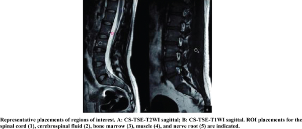
Methods: This retrospective study of lumbar vertebrae MRI included 600 patients; 300 patients received SENSE-TSE and 300 patients received CS-TSE. The SENSE acceleration factor was 1.4 for T1WI, 1.7 for T2WI, and 1.7 for PDWI. The CS total acceleration factor was 2.4, 3.6, 4.0, and 4.0 for T1WI, T2WI, PDWI sagittal, and T2WI transverse, respectively. The image quality of each MRI sequence was evaluated objectively by the signal-to-noise ratio (SNR) and contrast-to-noise ratio (CNR) and subjectively on a five-point scale. Two radiologists independently reviewed the MRI sequences of the 300 patients receiving CS-TSE, and their diagnostic consistency was evaluated. The degree of intervertebral foraminal stenosis and nerve root compression was assessed using the T1WI sagittal and T2WI transverse images.

Results: The scan time was reduced from 7 min 28 s to 4 min 26 s with CS-TSE. The median score of nerve root image quality was 5 (p > 0.05). The diagnostic consistency using CS-TSE images between the two radiologists was high for diagnosing lumbar diseases (κ > 0.75) and for evaluating the degree of lumbar foraminal stenosis and nerve root compression (κ = 0.882). No differences between SENSE-TSE and CS-TSE were observed for sensitivity, specificity, positive predictive value, or negative predictive value.

Conclusion: CS-TSE has the potential for diagnosing lumbar vertebrae and disc disorders.

Keywords: Lumbar vertebrae, nerve root, magnetic resonance imaging, compressed SENSE, turbo spin-echo, radiofrequency (RF).

Graphical Abstract
[1]
Goceri N, Goceri E. A Neural Network Based Kidney Segmentation from MR Images. Miami, Florida, USA: The 14th IEEE International Conference on Machine Learning and Applications. 1195-8.
[http://dx.doi.org/10.1109/ICMLA.2015.229]
[2]
Goceri E. Automatic Kidney Segmentation Using Gaussian Mixture Model on MRI Sequences. In: Wan X. (eds) Electrical Power Systems and Computers. Lecture Notes in Electrical Engineering, 2011; vol 99. Springer, Berlin, Heidelberg..
[http://dx.doi.org/10.1007/978-3-642-21747-0_4]
[3]
Goceri E. A comparative evaluation for liver segmentation from spir images and a novel level set method using signed pressure force function Izmir: Izmir Institute of Technology. 2013.
[4]
Goceri E, Songul C. Biomedical Information Technology: Image Based Computer Aided Diagnosis Systems. Antalya, Turkey Int Conf on Advanced Technologies.
[5]
Goceri E. Automatic labeling of portal and hepatic veins from MR images prior to liver transplantation. Int J CARS 2016; 11(12): 2153-61.
[http://dx.doi.org/10.1007/s11548-016-1446-8] [PMID: 27338273]
[6]
Goceri E, Shah ZK, Gurcan MN. Vessel segmentation from abdominal magnetic resonance images: adaptive and reconstructive approach. Int J Numer Methods Biomed Eng 2017; 33(4): Epub 2016 Aug 2.
[http://dx.doi.org/10.1002/cnm.2811] [PMID: 27315322]
[7]
Feng L, Benkert T, Block KT, Sodickson DK, Otazo R, Chandarana H. Compressed sensing for body MRI. J Magn Reson Imaging 2017; 45(4): 966-87.
[http://dx.doi.org/10.1002/jmri.25547] [PMID: 27981664]
[8]
Pillastrini P, Gardenghi I, Bonetti F, et al. An updated overview of clinical guidelines for chronic low back pain management in primary care. Joint Bone Spine 2012; 79(2): 176-85.
[http://dx.doi.org/10.1016/j.jbspin.2011.03.019] [PMID: 21565540]
[9]
Pruessmann KP, Weiger M, Scheidegger MB, Boesiger P. SENSE: sensitivity encoding for fast MRI. Magn Reson Med 1999; 42(5): 952-62.
[http://dx.doi.org/10.1002/(SICI)1522-2594(199911)42:5<952::AID-MRM16>3.0.CO;2-S] [PMID: 10542355]
[10]
David LD. Compressed sensing. IEEE Trans Inf 2006; 52: 1289-306.
[http://dx.doi.org/10.1109/TIT.2006.871582]
[11]
Lustig M, Donoho D, Pauly JM. Sparse MRI: The application of compressed sensing for rapid MR imaging. Magn Reson Med 2007; 58(6): 1182-95.
[http://dx.doi.org/10.1002/mrm.21391] [PMID: 17969013]
[12]
Jaspan ON, Fleysher R, Lipton ML. Compressed sensing MRI: a review of the clinical literature. Br J Radiol 2015; 88(1056): 20150487.
[http://dx.doi.org/10.1259/bjr.20150487] [PMID: 26402216]
[13]
Zhang T, Chowdhury S, Lustig M, et al. Clinical performance of contrast enhanced abdominal pediatric MRI with fast combined parallel imaging compressed sensing reconstruction. J Magn Reson Imaging 2014; 40(1): 13-25.
[http://dx.doi.org/10.1002/jmri.24333] [PMID: 24127123]
[14]
Liu F, Duan Y, Peterson BS, Kangarlu A. Compressed sensing MRI combined with SENSE in partial k-space. Phys Med Biol 2012; 57(21): N391-403.
[http://dx.doi.org/10.1088/0031-9155/57/21/N391] [PMID: 23073235]
[15]
Bratke G, Rau R, Weiss K, et al. Accelerated MRI of the Lumbar spine using compressed sensing: Quality and efficiency. J Magn Reson Imaging 2018 2019; 49(7): e164-75.
[PMID: 30267462]
[16]
Altahawi FF, Blount KJ, Morley NP, Raithel E, Omar IM. Comparing an accelerated 3D fast spin-echo sequence (CS-SPACE) for knee 3-T magnetic resonance imaging with traditional 3D fast spin-echo (SPACE) and routine 2D sequences. Skeletal Radiol 2017; 46(1): 7-15.
[http://dx.doi.org/10.1007/s00256-016-2490-8] [PMID: 27744578]
[17]
Yi J, Lee YH, Hahn S, Albakheet SS, Song H-T, Suh J-S. Fast isotropic volumetric magnetic resonance imaging of the ankle: Acceleration of the three-dimensional fast spin echo sequence using compressed sensing combined with parallel imaging. Eur J Radiol 2019; 112: 52-8.
[http://dx.doi.org/10.1016/j.ejrad.2019.01.009] [PMID: 30777219]
[18]
Kijowski R, Rosas H, Samsonov A, King K, Peters R, Liu F. Knee imaging: Rapid three-dimensional fast spin-echo using compressed sensing. J Magn Reson Imaging 2017; 45(6): 1712-22.
[http://dx.doi.org/10.1002/jmri.25507] [PMID: 27726244]
[19]
Lee SH, Lee YH, Song H-T, Suh J-S. Rapid acquisition of magnetic resonance imaging of the shoulder using three-dimensional fast spin echo sequence with compressed sensing. Magn Reson Imaging 2017; 42: 152-7.
[http://dx.doi.org/10.1016/j.mri.2017.07.022] [PMID: 28751204]
[20]
Uecker M, Lai P, Murphy MJ, et al. ESPIRiT- an eigenvalue approach to autocalibrating parallel MRI: where SENSE meets GRAPPA. Magn Reson Med 2014; 71(3): 990-1001.
[http://dx.doi.org/10.1002/mrm.24751] [PMID: 23649942]
[21]
Hu Y, Pan S, Zhao X, Guo W, He M, Guo Q. Value and clinical application of orthopedic metal artifact reduction algorithm in CT scans after orthopedic metal implantation. Korean J Radiol 2017; 18(3): 526-35.
[http://dx.doi.org/10.3348/kjr.2017.18.3.526] [PMID: 28458605]
[22]
Lee S, Lee JW, Yeom JS, et al. A practical MRI grading system for lumbar foraminal stenosis. AJR Am J Roentgenol 2010; 194(4): 1095-8.
[http://dx.doi.org/10.2214/AJR.09.2772] [PMID: 20308517]
[23]
Schizas C, Theumann N, Burn A, et al. Qualitative grading of severity of lumbar spinal stenosis based on the morphology of the dural sac on magnetic resonance images. Spine 2010; 35(21): 1919-24.
[http://dx.doi.org/10.1097/BRS.0b013e3181d359bd] [PMID: 20671589]
[24]
Xiong X, Zhou Z, Figini M, Shangguan J, Zhang Z, Chen W. Multi-parameter evaluation of lumbar intervertebral disc degeneration using quantitative magnetic resonance imaging techniques. Am J Transl Res 2018; 10(2): 444-54.
[PMID: 29511438]
[25]
Urrutia J, Besa P, Campos M, et al. The Pfirrmann classification of lumbar intervertebral disc degeneration: an independent inter- and intra-observer agreement assessment. Eur Spine J 2016; 25(9): 2728-33.
[http://dx.doi.org/10.1007/s00586-016-4438-z] [PMID: 26879918]
[26]
Fritz J, Raithel E, Thawait GK, Gilson W, Papp DF. Six-fold acceleration of high-spatial resolution 3D SPACE MRI of the knee through incoherent k-Space undersampling and iterative reconstruction-first experience. Invest Radiol 2016; 51(6): 400-9.
[http://dx.doi.org/10.1097/RLI.0000000000000240] [PMID: 26685106]
[27]
Delfaut EM, Beltran J, Johnson G, Rousseau J, Marchandise X, Cotten A. Fat suppression in MR imaging: techniques and pitfalls. Radiographics 1999; 19(2): 373-82.
[http://dx.doi.org/10.1148/radiographics.19.2.g99mr03373] [PMID: 10194785]
[28]
Lee S-Y, Jee W-H, Kim SK, Kim J-M. Proton density-weighted MR imaging of the knee: fat suppression versus without fat suppression. Skeletal Radiol 2011; 40(2): 189-95.
[http://dx.doi.org/10.1007/s00256-010-0969-2] [PMID: 20512570]
[29]
Hollingsworth KG. Reducing acquisition time in clinical MRI by data undersampling and compressed sensing reconstruction. Phys Med Biol 2015; 60(21): R297-322.
[http://dx.doi.org/10.1088/0031-9155/60/21/R297] [PMID: 26448064]
[30]
de Zwart JA, Ledden PJ, van Gelderen P, Bodurka J, Chu R, Duyn JH. Signal-to-noise ratio and parallel imaging performance of a 16-channel receive-only brain coil array at 3.0 Tesla. Magn Reson Med 2004; 51(1): 22-6.
[http://dx.doi.org/10.1002/mrm.10678] [PMID: 14705041]
[31]
Link TM. MR imaging in osteoarthritis: hardware, coils, and sequences. Radiol Clin North Am 2009; 47(4): 617-32.
[http://dx.doi.org/10.1016/j.rcl.2009.04.002] [PMID: 19631072]

© 2024 Bentham Science Publishers | Privacy Policy