Generic placeholder image

Current Medical Imaging

Editor-in-Chief

ISSN (Print): 1573-4056
ISSN (Online): 1875-6603

Mini-Review Article

Chest Radiograph (CXR) Manifestations of the Novel Coronavirus Disease 2019 (COVID-19): A Mini-review

Author(s): Wai Yee Chan*, Marlina Tanty Ramli Hamid*, Nadia Fareeda Muhammad Gowdh, Kartini Rahmat, Nur Adura Yaakup and Chee Shee Chai

Volume 17, Issue 6, 2021

Published on: 31 December, 2020

Page: [677 - 685] Pages: 9

DOI: 10.2174/1573405616666201231103312

Price: $65

Abstract

Background: Coronavirus disease 2019 (COVID-19) is highly contagious and has claimed more than one million lives, besides causing hardship and disruptions. The Fleischner Society has recommended chest X-ray (CXR) in detecting cases at high risk of disease progression, for triaging suspected patients with moderate-to-severe illness, and for eliminating false negatives in areas with high pre-test probability or limited resources. Although CXR is less sensitive than real-- time reverse transcription-polymerase chain reaction (RT-PCR) in detecting mild COVID-19, it is nevertheless useful because of equipment portability, low cost and practicality in serial assessments of disease progression among hospitalized patients.

Objective: This study aims to review the typical and relatively atypical CXR manifestations of COVID-19 pneumonia in a tertiary care hospital.

Methods: The CXRs of 136 COVID-19 patients confirmed through real-time RT-PCR from March to May 2020 were reviewed. A literature search was performed using PubMed.

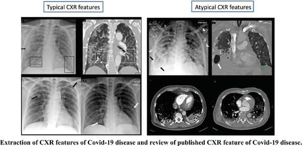
Results: A total of 54 patients had abnormal CXR whilst the others were normal. Typical CXR findings included pulmonary consolidation or ground-glass opacities in a multifocal, bilateral peripheral, or lower zone distribution, whereas atypical CXR features comprised cavitation and pleural effusion.

Conclusion: Typical findings of COVID-19 infection in chest computed tomography studies can also be seen in CXR. The presence of atypical features associated with worse disease outcome. Recognition of these features on CXR will improve the accuracy and speed of diagnosing COVID-19 patients.

Keywords: COVID-19, CXR, pneumonia, ground-glass opacity, consolidation, peripheral, ARDS.

Graphical Abstract
[1]
COVID-19 CORONAVIRUS PANDEMIC Worldometer 2020.https://www.worldometers.info/coronavirus/
[2]
Emergencies-Coronavirus disease (COVID-19) Pandemic World Health Organisation 2020.https://www.who.int/emergencies/diseases/novel-coronavirus-2019/events-as-they-happen/
[3]
Tang Y-W, Schmitz JE, Persing DH, Stratton CW. The laboratory diagnosis of COVID-19 infection: Current issues and challenges. J Clin Microbiol 2020; 58(6): e00512-20.
[http://dx.doi.org/10.1128/JCM.00512-20]
[4]
Sheridan C. Fast, portable tests come online to curb coronavirus pandemic. Nat Biotechnol 2020; 38(5): 515-8.
[http://dx.doi.org/10.1038/d41587-020-00010-2] [PMID: 32203294]
[5]
ACR Recommendations for the use of Chest Radiography and Computed Tomography (CT) for Suspected COVID-19 Infection American College Of Radiology 2020.https://www.acr.org/Advocacy-and-Economics/ACR-Position-Statements/Recommendations-for-Chest-Radiography-and-CT-for-Suspected-COVID19-Infection
[6]
Wong HYF, Lam HYS, Fong AH-T, et al. Frequency and distribution of chest radiographic findings in COVID-19 positive patients. Radiology 2020; 296(2): E72-8.
[http://dx.doi.org/10.1148/radiol.2020201160] [PMID: 32216717]
[7]
Rubin GD, Ryerson CJ, Haramati LB, et al. The role of chest imaging in patient management during the COVID-19 pandemic: A multinational consensus statement from the Fleischner Society. Chest 2020; 158(1): 106-16.
[http://dx.doi.org/10.1016/j.chest.2020.04.003] [PMID: 32275978]
[8]
Wu Z, McGoogan JM. Characteristics of and important lessons from the coronavirus disease 2019 (COVID-19) outbreak in China: summary of a report of 72 314 cases from the Chinese Center for Disease Control and Prevention. JAMA 2020; 323(13): 1239-42.
[http://dx.doi.org/10.1001/jama.2020.2648]
[9]
Organisation WH. Report of the WHO-China Joint Mission on Coronavirus Disease 2019 (COVID-19) 2020.https://www.who.int/docs/default-source/coronaviruse/who-china-joint-mission-on- covid-19-final-report.pdf
[10]
Guan WJ, Ni ZY, Hu Y, et al. China Medical Treatment Expert Group for Covid-19. Clinical characteristics of coronavirus disease 2019 in China. N Engl J Med 2020; 382(18): 1708-20.
[http://dx.doi.org/10.1056/NEJMoa2002032] [PMID: 32109013]
[11]
Chen N, Zhou M, Dong X, et al. Epidemiological and clinical characteristics of 99 cases of 2019 novel coronavirus pneumonia in Wuhan, China: a descriptive study. Lancet 2020; 395(10223): 507-13.
[http://dx.doi.org/10.1016/S0140-6736(20)30211-7] [PMID: 32007143]
[12]
Jin Y-H, Cai L, Cheng Z-S, et al. for the Zhongnan Hospital of Wuhan University Novel Coronavirus Management and Research Team, Evidence-Based Medicine Chapter of China International Exchange and Promotive Association for Medical and Health Care (CPAM). A rapid advice guideline for the diagnosis and treatment of 2019 novel coronavirus (2019-nCoV) infected pneumonia (standard version). Mil Med Res 2020; 7(1): 4.
[http://dx.doi.org/10.1186/s40779-020-0233-6] [PMID: 32029004]
[13]
Yoon SH, Lee KH, Kim JY, et al. Chest radiographic and CT findings of the 2019 novel coronavirus disease (COVID-19): Analysis of nine patients treated in Korea. Korean J Radiol 2020; 21(4): 494-500.
[http://dx.doi.org/10.3348/kjr.2020.0132] [PMID: 32100485]
[14]
Ng M-Y, Lee EY, Yang J, et al. Imaging profile of the COVID-19 infection: radiologic findings and literature review. Radiology. Cardiothoracic Imaging 2020; 2(1): 1-9.
[http://dx.doi.org/10.1148/ryct.2020200034]
[15]
Holshue ML, DeBolt C, Lindquist S, et al. Washington State 2019-nCoV Case Investigation Team. First case of 2019 novel coronavirus in the United States. N Engl J Med 2020; 382(10): 929-36.
[http://dx.doi.org/10.1056/NEJMoa2001191] [PMID: 32004427]
[16]
Salehi S, Abedi A, Balakrishnan S, Gholamrezanezhad A. Coronavirus disease 2019 (COVID-19): A systematic review of imaging findings in 919 patients. AJR Am J Roentgenol 2020; 215(1): 87-93.
[http://dx.doi.org/10.2214/AJR.20.23034] [PMID: 32174129]
[17]
Zhu N, Zhang D, Wang W, et al. China Novel Coronavirus Investigating and Research Team. A novel coronavirus from patients with pneumonia in China, 2019. N Engl J Med 2020; 382(8): 727-33.
[http://dx.doi.org/10.1056/NEJMoa2001017] [PMID: 31978945]
[18]
Rodrigues JCL, Hare SS, Edey A, et al. An update on COVID-19 for the radiologist - A British society of Thoracic Imaging statement. Clin Radiol 2020; 75(5): 323-5.
[http://dx.doi.org/10.1016/j.crad.2020.03.003] [PMID: 32216962]
[19]
Zu ZY, Jiang MD, Xu PP, et al. Coronavirus disease 2019 (COVID-19): A perspective from China. Radiology 2020; 296(2): E15-25.
[http://dx.doi.org/10.1148/radiol.2020200490] [PMID: 32083985]
[20]
Wang YXJ, Liu W-H, Yang M, Chen W. The role of CT for Covid-19 patient’s management remains poorly defined. Ann Transl Med 2020; 8(4): 145.
[http://dx.doi.org/10.21037/atm.2020.02.71] [PMID: 32175437]
[21]
Kanne JP, Little BP, Chung JH, Elicker BM, Ketai LH. Essentials for radiologists on COVID-19: an update—radiology scientific expert panel. Radiological Society of North America 2020.
[22]
Hosseiny M, Kooraki S, Gholamrezanezhad A, Reddy S, Myers L. Radiology perspective of coronavirus disease 2019 (COVID-19): lessons from severe acute respiratory syndrome and Middle East respiratory syndrome. AJR Am J Roentgenol 2020; 214(5): 1078-82.
[http://dx.doi.org/10.2214/AJR.20.22969] [PMID: 32108495]
[23]
Borghesi A, Zigliani A, Masciullo R, et al. Radiographic severity index in COVID-19 pneumonia: relationship to age and sex in 783 Italian patients. Radiol Med (Torino) 2020; 125(5): 461-4.
[http://dx.doi.org/10.1007/s11547-020-01202-1] [PMID: 32358691]
[24]
Schiaffino S, Tritella S, Cozzi A, et al. Diagnostic performance of chest X-Ray for COVID-19 pneumonia during the SARS-CoV-2 pandemic in Lombardy, Italy. J Thorac Imaging 2020; 35(4): W105-6.
[http://dx.doi.org/10.1097/RTI.0000000000000533] [PMID: 32404797]
[25]
Toussie D, Voutsinas N, Finkelstein M, et al. Clinical and chest radiography features determine patient outcomes in young and middle age adults with COVID-19. Radiology 2020; 297(1): E197-206.
[http://dx.doi.org/10.1148/radiol.2020201754] [PMID: 32407255]
[26]
Vancheri SG, Savietto G, Ballati F, et al. Radiographic findings in 240 patients with COVID-19 pneumonia: time-dependence after the onset of symptoms. Eur Radiol 2020; 30(11): 6161-9.
[http://dx.doi.org/10.1007/s00330-020-06967-7] [PMID: 32474630]
[27]
Hansell DM, Bankier AA, MacMahon H, McLoud TC, Müller NL, Remy J. Fleischner Society: glossary of terms for thoracic imaging. Radiology 2008; 246(3): 697-722.
[http://dx.doi.org/10.1148/radiol.2462070712] [PMID: 18195376]
[28]
Pan F, Ye T, Sun P, et al. Time course of lung changes on chest CT during recovery from 2019 novel coronavirus (COVID-19) pneumonia. Radiology 2020; 295(3): 715-21.
[http://dx.doi.org/10.1148/radiol.2020200370]
[29]
Xu Z, Shi L, Wang Y, et al. Pathological findings of COVID-19 associated with acute respiratory distress syndrome. Lancet Respir Med 2020; 8(4): 420-2.
[http://dx.doi.org/10.1016/S2213-2600(20)30076-X] [PMID: 32085846]
[30]
Ye Z, Zhang Y, Wang Y, Huang Z, Song B. Chest CT manifestations of new coronavirus disease 2019 (COVID-19): a pictorial review. Eur Radiol 2020; 30(8): 4381-9.
[http://dx.doi.org/10.1007/s00330-020-06801-0] [PMID: 32193638]
[31]
Song F, Shi N, Shan F, et al. Emerging 2019 novel coronavirus (2019-nCoV) pneumonia. Radiology 2020; 295(1): 210-7.
[http://dx.doi.org/10.1148/radiol.2020200274] [PMID: 32027573]
[32]
Shi H, Han X, Jiang N, et al. Radiological findings from 81 patients with COVID-19 pneumonia in Wuhan, China: a descriptive study. Lancet Infect Dis 2020; 20(4): 425-34.
[http://dx.doi.org/10.1016/S1473-3099(20)30086-4] [PMID: 32105637]
[33]
Bernheim A, Mei X, Huang M, et al. Chest CT findings in coronavirus disease-19 (COVID-19): Relationship to duration of infection. Radiology 2020; 295(3): 200463.
[http://dx.doi.org/10.1148/radiol.2020200463] [PMID: 32077789]
[34]
Wu C, Chen X, Cai Y, et al. Risk factors associated with acute respiratory distress syndrome and death in patients with coronavirus disease 2019 pneumonia in Wuhan, China. JAMA Intern Med 2020; 180(7): 934-43.
[http://dx.doi.org/10.1001/jamainternmed.2020.0994] [PMID: 32167524]
[35]
Ai T, Yang Z, Hou H, et al. Correlation of chest CT and RT-PCR testing in coronavirus disease 2019 (COVID-19) in China: A report of 1014 cases. Radiology 2020; 296(2): E32-40.
[http://dx.doi.org/10.1148/radiol.2020200642] [PMID: 32101510]
[36]
Kanne JP. Chest CT findings in 2019 novel coronavirus (2019-nCoV) infections from Wuhan, China: key points for the radiologist. Radiology 2020; 295(1): 16-7.
[http://dx.doi.org/10.1148/radiol.2020200241] [PMID: 32017662]
[37]
Xie X, Zhong Z, Zhao W, Zheng C, Wang F, Liu J. Chest CT for typical 2019-nCoV pneumonia: relationship to negative RT-PCR testing. Radiology 2020; 296(2): E41-5.
[38]
Aviram G, Bar-Shai A, Sosna J, et al. H1N1 influenza: initial chest radiographic findings in helping predict patient outcome. Radiology 2010; 255(1): 252-9.
[http://dx.doi.org/10.1148/radiol.10092240] [PMID: 20308461]
[39]
Chung M, Bernheim A, Mei X, et al. CT imaging features of 2019 novel coronavirus (2019-nCoV). Radiology 2020; 295(1): 202-7.
[http://dx.doi.org/10.1148/radiol.2020200230] [PMID: 32017661]
[40]
Jacobi A, Chung M, Bernheim A, Eber C. Portable chest X-ray in coronavirus disease-19 (COVID-19): A pictorial review. Clin Imaging 2020; 64: 35-42.
[http://dx.doi.org/10.1016/j.clinimag.2020.04.001] [PMID: 32302927]
[41]
Li K, Wu J, Wu F, et al. The clinical and chest CT features associated with severe and critical COVID-19 pneumonia. Invest Radiol 2020; 55(6): 327-31.
[http://dx.doi.org/10.1097/RLI.0000000000000672] [PMID: 32118615]
[42]
Gibbs JM, Chandrasekhar CA, Ferguson EC, Oldham SA. Lines and stripes: where did they go?--From conventional radiography to CT. Radiographics 2007; 27(1): 33-48.
[http://dx.doi.org/10.1148/rg.271065073] [PMID: 17234997]
[43]
Co SJ, Yong-Hing CJ, Galea-Soler S, et al. Role of imaging in penetrating and blunt traumatic injury to the heart. Radiographics 2011; 31(4): E101-15.
[http://dx.doi.org/10.1148/rg.314095177] [PMID: 21768229]

Rights & Permissions Print Cite
© 2024 Bentham Science Publishers | Privacy Policy