Generic placeholder image

Current Medical Imaging

Editor-in-Chief

ISSN (Print): 1573-4056
ISSN (Online): 1875-6603

Research Article

Computer Simulation of the Effects of Contrast Protocols on Aortic Signal Intensity on Magnetic Resonance Angiograms

Author(s): Toru Higaki*, Yuko Nakamura, Fuminari Tatsugami, Wataru Fukumoto and Kazuo Awai

Volume 17, Issue 3, 2021

Published on: 30 July, 2020

Page: [396 - 403] Pages: 8

DOI: 10.2174/1573405616999200730180533

Price: $65

Abstract

Background: While iodine-enhanced computed tomography has been studied, detailed information on gadolinium-enhanced magnetic resonance imaging has not been reported.

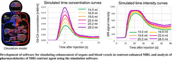
Objective: We evaluated the effects of different gadolinium contrast agent (Gd-CA) factors on the enhancement of aortic magnetic resonance angiography (MRA) using computer simulation.

Methods: We developed computer-simulation software that combines pharmacokinetic models and tables; it converts the blood concentration of particular Gd-CAs into the signal intensity (SI). We simulated aortic time-intensity curves (TIC) in our MRA study and compared the effect of the Gd-- CA volume, injection rate, and of different Gd-CAs on the TIC.

Results: An increase in the Gd-CA volume from 14.0 to 28.0 ml increased maximal aortic intensity 1.11 times. Changing the injection rate from 1.0 to 2.8 ml/s increased it 1.10 times. The maximal SI of gadoterate-meglumine and gadobutrol was 1.03 and 1.01 times, respectively, that of gadoteridol.

Conclusion: In our computer-simulated MRA study, different Gd-CA factors resulted in no significant difference in the maximal aortic SI.

Keywords: Gadolinium contrast agent, contrast-enhanced MR, MR angiography, computer simulation, contrast protocol, time-intensity curve.

Graphical Abstract
[1]
Gerretsen SC, le Maire TF, Miller S, et al. Multicenter, double-blind, randomized, intraindividual crossover comparison of gadobenate dimeglumine and gadopentetate dimeglumine for MR angiography of peripheral arteries. Radiology 2010; 255(3): 988-1000.
[http://dx.doi.org/10.1148/radiol.10090357] [PMID: 20501735]
[2]
Haneder S, Attenberger UI, Schoenberg SO, Loewe C, Arnaiz J, Michaely HJ. Comparison of 0.5 M gadoterate and 1.0 M gadobutrol in peripheral MRA: a prospective, single-center, randomized, crossover, double-blind study. J Magn Reson Imaging 2012; 36(5): 1213-21.
[http://dx.doi.org/10.1002/jmri.23760] [PMID: 22848033]
[3]
Hrdina L, Kocher M, Herman M, et al. Comparison of the quality of lower limb magnetic resonance angiographies performed with different paramagnetic contrast agents in relation to body mass index and ejection fraction. Biomed Pap Med Fac Univ Palacky Olomouc Czech Repub 2012; 156(2): 164-70.
[http://dx.doi.org/10.5507/bp.2011.058] [PMID: 22660207]
[4]
Kramer JH, Arnoldi E, François CJ, et al. Dynamic and static magnetic resonance angiography of the supra-aortic vessels at 3.0 T: intraindividual comparison of gadobutrol, gadobenate dimeglumine, and gadoterate meglumine at equimolar dose. Invest Radiol 2013; 48(3): 121-8.
[http://dx.doi.org/10.1097/RLI.0b013e31827752b4] [PMID: 23211552]
[5]
Wang J, Yan F, Liu J, et al. Multicenter, intra-individual comparison of single dose gadobenate dimeglumine and double dose gadopentetate dimeglumine for MR angiography of the peripheral arteries (the Peripheral VALUE Study). J Magn Reson Imaging 2013; 38(4): 926-37.
[http://dx.doi.org/10.1002/jmri.24040] [PMID: 23371919]
[6]
Hansmann J, Michaely HJ, Morelli JN, Luckscheiter A, Schoenberg SO, Attenberger UI. Enhancement characteristics and impact on image quality of two gadolinium chelates at equimolar doses for time-resolved 3-Tesla MR-angiography of the calf station. PLoS One 2014; 9(6)e99079
[http://dx.doi.org/10.1371/journal.pone.0099079] [PMID: 24893292]
[7]
Loewe C, Arnaiz J, Krause D, Marti-Bonmati L, Haneder S, Kramer U. DALIA Study Group. MR angiography at 3 T of peripheral arterial disease: A randomized prospective comparison of gadoterate meglumine and gadobutrol. AJR Am J Roentgenol 2015; 204(6): 1311-21.
[http://dx.doi.org/10.2214/AJR.14.12604] [PMID: 26001243]
[8]
Hoelter P, Lang S, Weibart M, et al. Prospective intraindividual comparison of gadoterate and gadobutrol for cervical and intracranial contrast-enhanced magnetic resonance angiography. Neuroradiology 2017; 59(12): 1233-9.
[http://dx.doi.org/10.1007/s00234-017-1922-z] [PMID: 28913611]
[9]
Xing X, Zeng X, Li X, et al. Contrast-enhanced MR angiography: does a higher relaxivity MR contrast agent permit a reduction of the dose administered for routine vascular imaging applications? Radiol Med (Torino) 2015; 120(2): 239-50.
[http://dx.doi.org/10.1007/s11547-014-0434-8] [PMID: 25183340]
[10]
Fink C, Bock M, Kiessling F, et al. Time-resolved contrast-enhanced three-dimensional pulmonary MR-angiography: 1.0 M gadobutrol vs. 0.5 M gadopentetate dimeglumine. J Magn Reson Imaging 2004; 19(2): 202-8.
[http://dx.doi.org/10.1002/jmri.10452] [PMID: 14745754]
[11]
Fink C, Puderbach M, Ley S, et al. Contrast-enhanced three-dimensional pulmonary perfusion magnetic resonance imaging: intraindividual comparison of 1.0 M gadobutrol and 0.5 M Gd-DTPA at three dose levels. Invest Radiol 2004; 39(3): 143-8.
[http://dx.doi.org/10.1097/01.rli.0000101482.79137.f4] [PMID: 15076006]
[12]
Fink C, Puderbach M, Ley S, et al. Intraindividual comparison of 1.0 M gadobutrol and 0.5 M gadopentetate dimeglumine for time-resolved contrast-enhanced three-dimensional magnetic resonance angiography of the upper torso. J Magn Reson Imaging 2005; 22(2): 286-90.
[http://dx.doi.org/10.1002/jmri.20381] [PMID: 16028246]
[13]
Carroll TJ, Korosec FR, Swan JS, Hany TF, Grist TM, Mistretta CA. The effect of injection rate on time-resolved contrast-enhanced peripheral MRA. J Magn Reson Imaging 2001; 14(4): 401-10.
[http://dx.doi.org/10.1002/jmri.1200] [PMID: 11599064]
[14]
Herold T, Paetzel C, Völk M, et al. Contrast-enhanced magnetic resonance angiography of the carotid arteries: influence of injection rates and volumes on arterial-venous transit time. Invest Radiol 2004; 39(2): 65-72.
[http://dx.doi.org/10.1097/01.rli.0000105040.40925.c8] [PMID: 14734920]
[15]
Kopka L, Vosshenrich R, Rodenwaldt J, Grabbe E. Differences in injection rates on contrast-enhanced breath-hold three-dimensional MR angiography. AJR Am J Roentgenol 1998; 170(2): 345-8.
[http://dx.doi.org/10.2214/ajr.170.2.9456943] [PMID: 9456943]
[16]
Kramer H, Michaely HJ, Requardt M, et al. Effects of injection rate and dose on image quality in time-resolved magnetic resonance angiography (MRA) by using 1.0M contrast agents. Eur Radiol 2007; 17(6): 1394-402.
[http://dx.doi.org/10.1007/s00330-006-0493-x] [PMID: 17115161]
[17]
Pannetier NA, Debacker CS, Mauconduit F, Christen T, Barbier EL. A simulation tool for dynamic contrast enhanced MRI. PLoS One 2013; 8(3)e57636
[http://dx.doi.org/10.1371/journal.pone.0057636] [PMID: 23516414]
[18]
Tofts PS. Modeling tracer kinetics in dynamic Gd-DTPA MR imaging. J Magn Reson Imaging 1997; 7(1): 91-101.
[http://dx.doi.org/10.1002/jmri.1880070113] [PMID: 9039598]
[19]
Bae KT, Heiken JP, Brink JA. Aortic and hepatic contrast medium enhancement at CT. Part II. Effect of reduced cardiac output in a porcine model. Radiology 1998; 207(3): 657-62.
[http://dx.doi.org/10.1148/radiology.207.3.9609887] [PMID: 9609887]
[20]
Bae KT, Heiken JP, Brink JA. Aortic and hepatic peak enhancement at CT: effect of contrast medium injection rate--pharmacokinetic analysis and experimental porcine model. Radiology 1998; 206(2): 455-64.
[http://dx.doi.org/10.1148/radiology.206.2.9457200] [PMID: 9457200]
[21]
Bae KT, Heiken JP, Brink JA. Aortic and hepatic contrast medium enhancement at CT. Part I. Prediction with a computer model. Radiology 1998; 207(3): 647-55.
[http://dx.doi.org/10.1148/radiology.207.3.9609886] [PMID: 9609886]
[22]
Higaki T, Nakaura T, Kidoh M, et al. Effect of contrast material injection duration on arterial enhancement at CT in patients with various cardiac indices: Analysis using computer simulation. PLoS One 2018; 13(2)e0191347
[http://dx.doi.org/10.1371/journal.pone.0191347] [PMID: 29474457]
[23]
Sahbaee P, Segars WP, Marin D, Nelson RC, Samei E. The effect of contrast material on radiation dose at CT: Part I. Incorporation of contrast material dynamics in anthropomorphic phantoms. Radiology 2017; 283(3): 739-48.
[http://dx.doi.org/10.1148/radiol.2016152851] [PMID: 28092496]
[24]
Bae KT. Peak contrast enhancement in CT and MR angiography: when does it occur and why? Pharmacokinetic study in a porcine model. Radiology 2003; 227(3): 809-16.
[http://dx.doi.org/10.1148/radiol.2273020102] [PMID: 12702823]
[25]
Bae KT. Intravenous contrast medium administration and scan timing at CT: considerations and approaches. Radiology 2010; 256(1): 32-61.
[http://dx.doi.org/10.1148/radiol.10090908] [PMID: 20574084]
[26]
Bae KT. Optimization of contrast enhancement in thoracic MDCT. Radiol Clin North Am 2010; 48(1): 9-29.
[http://dx.doi.org/10.1016/j.rcl.2009.08.012] [PMID: 19995627]
[27]
Golkov V, Dosovitskiy A, Sperl JI, et al. q-Space deep learning: Twelve-fold shorter and model-free diffusion MRI scans. IEEE Trans Med Imaging 2016; 35(5): 1344-51.
[http://dx.doi.org/10.1109/TMI.2016.2551324] [PMID: 27071165]
[28]
Awai K, Nakayama Y, Nakaura T, et al. Prediction of aortic peak enhancement in monophasic contrast injection protocols at multidetector CT: phantom and patient studies. Radiat Med 2007; 25(1): 14-21.
[http://dx.doi.org/10.1007/s11604-006-0095-1] [PMID: 17225048]
[29]
Rohrer M, Bauer H, Mintorovitch J, Requardt M, Weinmann HJ. Comparison of magnetic properties of MRI contrast media solutions at different magnetic field strengths. Invest Radiol 2005; 40(11): 715-24.
[http://dx.doi.org/10.1097/01.rli.0000184756.66360.d3] [PMID: 16230904]
[30]
Essig M, Lodemann KP, Le-Huu M, Brüning R, Kirchin M, Reith W. Intraindividual comparison of gadobenate dimeglumine and gadobutrol for cerebral magnetic resonance perfusion imaging at 1.5 T. Invest Radiol 2006; 41(3): 256-63.
[http://dx.doi.org/10.1097/01.rli.0000191333.19068.6b] [PMID: 16481908]
[31]
Tombach B, Benner T, Reimer P, et al. Do highly concentrated gadolinium chelates improve MR brain perfusion imaging? Intraindividually controlled randomized crossover concentration comparison study of 0.5 versus 1.0 mol/L gadobutrol. Radiology 2003; 226(3): 880-8.
[http://dx.doi.org/10.1148/radiol.2263011970] [PMID: 12601217]
[32]
Cruite I, Schroeder M, Merkle EM, Sirlin CB. Gadoxetate disodium-enhanced MRI of the liver: part 2, protocol optimization and lesion appearance in the cirrhotic liver. AJR Am J Roentgenol 2010; 195(1): 29-41.
[http://dx.doi.org/10.2214/AJR.10.4538] [PMID: 20566795]
[33]
Ringe KI, Husarik DB, Sirlin CB, Merkle EM. Gadoxetate disodium-enhanced MRI of the liver: part 1, protocol optimization and lesion appearance in the noncirrhotic liver. AJR Am J Roentgenol 2010; 195(1): 13-28.
[http://dx.doi.org/10.2214/AJR.10.4392] [PMID: 20566794]

Rights & Permissions Print Cite
© 2024 Bentham Science Publishers | Privacy Policy