Generic placeholder image

Current Medical Imaging

Editor-in-Chief

ISSN (Print): 1573-4056
ISSN (Online): 1875-6603

Research Article

Radiologic Findings of a Rare Subtype of Invasive Breast Cancer with Poor Prognosis: Metaplastic Carcinoma of the Breast

Author(s): Hale Aydin*, Bahar Guner, Isil Esen Bostanci, Nazan Ciledag, Melda Boyacioglu Bulut, Mustafa Bozgul, Gulay Bilir Dilek and Bilgin Kadri Aribas

Volume 15, Issue 3, 2019

Page: [338 - 348] Pages: 11

DOI: 10.2174/1573405614666180813120227

Price: $65

Abstract

Background and Objectives: The purpose of this study was to evaluate the mammographic, sonographic and MRI findings of metaplastic breast carcinoma.

Methods: In this retrospective review study, we analyzed the medical files of 9600 patients who were treated for invasive breast cancers. Clinical information, histopathologic and radiologic findings of 65 patients were included in this study. All existing radiologic images and medical reports were reviewed retrospectively. Thirty-three patients had MG, 58 patients had US and 7 patients had MRI imaging results.

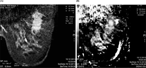
Results: Mammographically, the most frequent presentations of MPBC were round shape, microlobulated margin and high density masses. Calcifications with or without masses were not a frequent finding. The most common sonographic findings were round shape, partially indistinct angular margin, hypoechoic and heterogeneous echo patterns and no posterior feature masses. All lesions were presented as masses rather than non-mass enhancements on magnetic resonance imaging. Features of masses had more malignant feature on MRI than other modalities in all 7 patients.

Conclusion: Metaplastic breast carcinoma is one of the rarest poorly differentiated invasive breast carcinomas. Interestingly, these aggressive tumors demonstrate benign or moderately malign features on imaging methods. This appearance of MPBC can cause it to be misdiagnosed as a benign breast lesion especially in young women. MPBC should be kept in mind in the differential diagnosis of large palpable breast masses. Therefore, follow-up at short intervals and/or multimodality imaging studies which include breast MRI are important for the diagnosis of MPBC.

Keywords: Metaplastic breast carcinoma, mammography, ultrasonography, MRI, patients, malignant.

Graphical Abstract
[1]
Luini A, Aguilar M, Gatti G, et al. Metaplastic carcinoma of the breast, an unusual disease with worse prognosis: The experience of the European Institute of Oncology and review of the literature. Breast Cancer Res Treat 2007; 101: 349-53.
[2]
Smith D, Rongaus V, Wehmann T, Agarwal P, Classen G. Metaplastic breast carcinoma. J Am Osteopath Assoc 1996; 96: 419-21.
[3]
Günhan-Bilgen Ii, Memis A, Üstün EE, Zekioglu O, Özdemir N. Metaplastic carcinoma of the breast: Clinical, mammographic, and sonographic findings with histopathologic correlation. AJR Am J Roentgenol 2002; 178: 1421-5.
[4]
Fritz A, Percy C, Jack A, et al. International classification of diseases for oncology: World Health Organization, 2000. Aavailable from: http://codes.iarc.fr/abouticdo.php
[5]
Lakhani SR, Ellis IO, Schnitt SJ, Tan PH, van de Vijver MJ. WHO classification of tumors of the breast: International agency for research on cancer. 4th ed. 2012.
[6]
Jung S-Y, Kim HY, Nam B-H, et al. Worse prognosis of metaplastic breast cancer patients than other patients with triple-negative breast cancer. Breast Cancer Res Treat 2010; 120: 627-37.
[7]
Okada N, Hasebe T, Iwasaki M, et al. Metaplastic carcinoma of the breast. Hum Pathol 2010; 41: 960-70.
[8]
Dieci MV, Orvieto E, Dominici M, Conte P, Guarneri V. Rare breast cancer subtypes: Histological, molecular, and clinical peculiarities. Oncologist 2014; 19: 805-13.
[9]
Schwartz TL, Mogal H, Papageorgiou C, Veerapong J, Hsueh EC. Metaplastic breast cancer: Histologic characteristics, prognostic factors and systemic treatment strategies. Exp Hematol Oncol 2013; 2: 31.
[10]
Yilmaz K, Pak I, Irkkan C, Ozaslan C, Atalay C. Metaplastic carcinoma of the breast: Clinicopathological features and immunohistochemical analysis. J BUON 2011; 16: 652-6.
[11]
Pezzi CM, Patel-Parekh L, Cole K, Franko J, Klimberg VS, Bland K. Characteristics and treatment of metaplastic breast cancer: Analysis of 892 cases from the National Cancer Data Base. Ann Surg Oncol 2007; 14: 166-73.
[12]
Karan B, Pourbagher A, Bolat FA. Unusual malignant breast lesions: imaging-pathological correlations. Diagn Interv Radiol 2012; 18: 270.
[13]
Patterson SK, Tworek JA, Roubidoux MA, Helvie MA, Oberman HA. Metaplastic carcinoma of the breast: Mammographic appearance with pathologic correlation. AJR Am J Roentgenol 1997; 169: 709-12.
[14]
Yang WT, Hennessy B, Broglio K, et al. Imaging differences in metaplastic and invasive ductal carcinomas of the breast. AJR Am J Roentgenol 2007; 189: 1288-93.
[15]
D’Orsi CJ, Sickles EA, Mendelson EB, et al. ACR BI-RADS Atlas, Breast Imaging Reporting and Data System. Reston, VA: Am Coll Radiol 2013.
[16]
Choi BB, Shu KS. Metaplastic carcinoma of the breast: Multimodality imaging and histopathologic assessment. Acta Radiol 2012; 53: 5-11.
[17]
Lai Y-C, Hsu C-Y, Chou Y-H, et al. Sonographic presentations of metaplastic breast cancers. J Chin Med Assoc 2012; 75: 589-94.
[18]
Velasco M, Santamaría G, Ganau S, et al. MRI of metaplastic carcinoma of the breast. Am J Roentgenol 2005; 184: 1274-8.
[19]
Yoo JL, Woo OH, Kim YK, et al. Can MR imaging contribute in characterizing well-circumscribed breast carcinomas? Radiographics 2010; 30: 1689-704.
[20]
Leddy R, Irshad A, Rumboldt T, Cluver A, Campbell A, Ackerman S. Review of metaplastic carcinoma of the breast: imaging findings and pathologic features. J Clin Imaging Sci 2012; 2: 21.
[21]
Iyengar P, Cody H, Brogi E. Pleomorphic adenoma of the breast: case report and review of the literature. Diagn Cytopathol 2005; 33: 416-20.
[22]
Tan PH, Ellis IO. Myoepithelial and epithelial–myoepithelial, mesenchymal and fibroepithelial breast lesions: updates from the WHO classification of tumors of the breast 2012. J Clin Pathol 2013; 66(6): 465-70.
[23]
Chang YW, Lee M, Kwon K, et al. Magnetic resonance imaging of metaplastic carcinoma of the breast: Sonographic and pathologic correlation. Acta Radiol 2004; 45: 18-22.
[24]
Evans H, Shaughnessy E, Nikiforov YE. Infiltrating ductal carcinoma of the breast with osseous metaplasia: Imaging findings with pathologic correlation. AJR Am J Roentgenol 1999; 172: 1420-2.
[25]
Park JM, Han BK, Moon WK, Choe YH, Ahn SH, Gong G. Metaplastic carcinoma of the breast: Mammographic and sonographic findings. J Clin Ultrasound 2000; 28: 179-86.
[26]
Shin HJ, Kim HH, Kim SM, et al. Imaging features of metaplastic carcinoma with chondroid differentiation of the breast. AJR Am J Roentgenol 2007; 188: 691-6.
[27]
M Lu, D Wu, W Lin, W Li, H Zhang, WH Huang. A stochastic filtering approach to recover strain images from quasi-static ultrasound elastography. Biomed Eng Online 2014; 13(1): 15.
[28]
M Guo, M Lu, H Zhang, H Liu, YT Zhang. A rod-like acoustic radiation force in ultrasound-based elastography: A simulation study. Zhang YT. (eds). In: The ınternational conference on health ınformatics, 2014; pp. 148-51.
[29]
Z Hu, H Zhang, J Yuan, M Lu, S Chen, H Liu. An H∞ strategy for strain estimation in ultrasound elastography using biomechanical modeling constraint. PLoS One 2013; 8(9): e73093.
[30]
M Lu, H Zhang, J Wang, J Yuan, Z Hu, H Liu. Reconstruction of elasticity: A stochastic model-based approach in ultrasound elastography. Biomed Eng Online 2013; 12(1): 79.
[31]
Evans A, Sim YT, Thomson K, Jordan L, Purdie C, Vinnicombe SJ. Shear wave elastography of breast cancer: Sensitivity according to histological type in a large cohort. Breast 2016; 26: 115-8.

Rights & Permissions Print Cite
© 2024 Bentham Science Publishers | Privacy Policy