Generic placeholder image

Current Medical Imaging

Editor-in-Chief

ISSN (Print): 1573-4056
ISSN (Online): 1875-6603

Research Article

Diffusion Tensor Imaging of Brain Metastases in Patients with Breast Cancer According to Molecular Subtypes

Author(s): Ismail Yurtsever*, Lutfullah Sari, Mehmet Ali Gultekin, Huseyin Toprak, Haci Mehmet Turk, Altay Aliyev, Abdusselim Adil Peker, Aysegul Yabaci and Alpay Alkan

Volume 17, Issue 1, 2021

Published on: 21 June, 2020

Page: [120 - 128] Pages: 9

DOI: 10.2174/1573405616666200621195655

Price: $65

Abstract

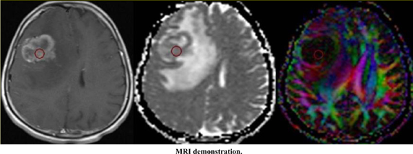
Background and Purpose: Recent studies have shown that diffusion tensor imaging (DTI) parameters are used to follow the patients with breast cancer and correlate well as a prognostic parameter of breast cancer. However, as far as we know, there is no data to compare the DTI features of breast cancer brain metastases according to molecular subtypes in the literature. Our aim is to evaluate whether there are any differences in DTI parameters of brain metastases in patients with breast cancer according to molecular subtypes.

Methods: Twenty-seven patients with breast cancer and 82 metastatic brain lesions were included. We classified subjects into three subgroups according to their hormone expression; Group 0, triple- negative (n; 6, 19 lesions), group 1, HER2-positive (n;16, 54 lesions) and group 2, hormone-- positive group (n; 5, 9 lesions). The apparent diffusion coefficient (ADC), fractional anisotropy (FA), axial diffusivity (AD), and radial diffusivity (RD) values in DTI were measured and compared between three groups.

Results: ADC, AD and RD values of group 2 were significantly lower compared to group 0. No significant differences were found in FA, ADC, AD and RD values between the group 0 and 1 and the group 1 and 2.

Conclusion: Metastasis of aggressive triple-negative breast cancer showed higher ADC values compared to the less aggressive hormone-positive group. Higher ADC values in brain metastases of breast cancer may indicate a poor prognosis, so DTI findings could play a role in planning appropriate treatment.

Keywords: Breast cancer, molecular subgroup, brain metastases, diffusion tensor imaging, fractional anisotropy (FA), axial diffusivity (AD), radial diffusivity (RD).

Graphical Abstract
[1]
Loo CE, Straver ME, Rodenhuis S, et al. Magnetic resonance imaging response monitoring of breast cancer during neoadjuvant chemotherapy: relevance of breast cancer subtype. J Clin Oncol 2011; 29(6): 660-6.
[http://dx.doi.org/10.1200/JCO.2010.31.1258] [PMID: 21220595]
[2]
Heitz F, Harter P, Lueck H-J, et al. Triple-negative and HER2-overexpressing breast cancers exhibit an elevated risk and an earlier occurrence of cerebral metastases. Eur J Cancer 2009; 45(16): 2792-8.
[http://dx.doi.org/10.1016/j.ejca.2009.06.027] [PMID: 19643597]
[3]
Tsukada Y, Fouad A, Pickren JW, Lane WW. Central nervous system metastasis from breast carcinoma. Autopsy study. Cancer 1983; 52(12): 2349-54.
[http://dx.doi.org/10.1002/1097-0142(19831215)52:12<2349::AID-CNCR2820521231>3.0.CO;2-B] [PMID: 6640506]
[4]
Cadoo KA, Fornier MN, Morris PG. Biological subtypes of breast cancer: current concepts and implications for recurrence patterns. Q J Nucl Med Mol Imaging 2013; 57(4): 312-21.
[PMID: 24322788]
[5]
Abdel Razek AAK, Zaky M, Bayoumi D, Taman S, Abdelwahab K, Alghandour R. Diffusion tensor imaging parameters in differentiation recurrent breast cancer from post-operative changes in patients with breast-conserving surgery. Eur J Radiol 2019; 111: 76-80.
[http://dx.doi.org/10.1016/j.ejrad.2018.12.022] [PMID: 30691669]
[6]
Aurilio G, Disalvatore D, Pruneri G, et al. A meta-analysis of oestrogen receptor, progesterone receptor and human epidermal growth factor receptor 2 discordance between primary breast cancer and metastases. Eur J Cancer 2014; 50(2): 277-89.
[http://dx.doi.org/10.1016/j.ejca.2013.10.004] [PMID: 24269135]
[7]
Abdel Razek AAK, El-Serougy L, Abdelsalam M, Gaballa G, Talaat M. Differentiation of primary central nervous system lymphoma from glioblastoma: Quantitative analysis using arterial spin labeling and diffusion tensor imaging. World Neurosurg 2019; 123: e303-9.
[http://dx.doi.org/10.1016/j.wneu.2018.11.155] [PMID: 30502475]
[8]
Liu S, Ren R, Chen Z, et al. Diffusion-weighted imaging in assessing pathological response of tumor in breast cancer subtype to neoadjuvant chemotherapy. J Magn Reson Imaging 2015; 42(3): 779-87.
[http://dx.doi.org/10.1002/jmri.24843] [PMID: 25580585]
[9]
El-Serougy L, Abdel Razek AA, Ezzat A, Eldawoody H, El-Morsy A. Assessment of diffusion tensor imaging metrics in differentiating low-grade from high-grade gliomas. Neuroradiol J 2016; 29(5): 400-7.
[http://dx.doi.org/10.1177/1971400916665382] [PMID: 27562582]
[10]
Razek AAKA, El-Serougy L, Abdelsalam M, Gaballa G, Talaat M. Differentiation of residual/recurrent gliomas from postradiation necrosis with arterial spin labeling and diffusion tensor magnetic resonance imaging-derived metrics. Neuroradiology 2018; 60(2): 169-77.
[http://dx.doi.org/10.1007/s00234-017-1955-3] [PMID: 29218370]
[11]
Guo Y, Cai YQ, Cai ZL, et al. Differentiation of clinically benign and malignant breast lesions using diffusion-weighted imaging. J Magn Reson Imaging 2002; 16(2): 172-8.
[http://dx.doi.org/10.1002/jmri.10140] [PMID: 12203765]
[12]
Woodhams R, Matsunaga K, Iwabuchi K, et al. Diffusion-weighted imaging of malignant breast tumors: the usefulness of apparent diffusion coefficient (ADC) value and ADC map for the detection of malignant breast tumors and evaluation of cancer extension. J Comput Assist Tomogr 2005; 29(5): 644-9.
[http://dx.doi.org/10.1097/01.rct.0000171913.74086.1b] [PMID: 16163035]
[13]
Yoshikawa MI, Ohsumi S, Sugata S, et al. Relation between cancer cellularity and apparent diffusion coefficient values using diffusion-weighted magnetic resonance imaging in breast cancer. Radiat Med 2008; 26(4): 222-6.
[http://dx.doi.org/10.1007/s11604-007-0218-3] [PMID: 18509722]
[14]
Partridge SC, Mullins CD, Kurland BF, et al. Apparent diffusion coefficient values for discriminating benign and malignant breast MRI lesions: effects of lesion type and size. AJR Am J Roentgenol 2010; 194(6): 1664-73.
[http://dx.doi.org/10.2214/AJR.09.3534] [PMID: 20489111]
[15]
Jeh SK, Kim SH, Kim HS, et al. Correlation of the apparent diffusion coefficient value and dynamic magnetic resonance imaging findings with prognostic factors in invasive ductal carcinoma. J Magn Reson Imaging 2011; 33(1): 102-9.
[http://dx.doi.org/10.1002/jmri.22400] [PMID: 21182127]
[16]
Chikarmane SA, Tirumani SH, Howard SA, Jagannathan JP, DiPiro PJ. Metastatic patterns of breast cancer subtypes: what radiologists should know in the era of personalized cancer medicine. Clin Radiol 2015; 70(1): 1-10.
[http://dx.doi.org/10.1016/j.crad.2014.08.015] [PMID: 25300558]
[17]
Yonemori K, Tsuta K, Ono M, et al. Disruption of the blood brain barrier by brain metastases of triple-negative and basal-type breast cancer but not HER2/neu-positive breast cancer. Cancer 2010; 116(2): 302-8.
[http://dx.doi.org/10.1002/cncr.24735] [PMID: 19937674]
[18]
Kamitani T, Matsuo Y, Yabuuchi H, et al. Correlations between apparent diffusion coefficient values and prognostic factors of breast cancer. Magn Reson Med Sci 2013; 12(3): 193-9.
[http://dx.doi.org/10.2463/mrms.2012-0095] [PMID: 23857151]
[19]
Kim EJ, Kim SH, Park GE, et al. Histogram analysis of apparent diffusion coefficient at 3.0t: Correlation with prognostic factors and subtypes of invasive ductal carcinoma. J Magn Reson Imaging 2015; 42(6): 1666-78.
[http://dx.doi.org/10.1002/jmri.24934] [PMID: 25919239]
[20]
Leek RD, Landers RJ, Harris AL, Lewis CE. Necrosis correlates with high vascular density and focal macrophage infiltration in invasive carcinoma of the breast. Br J Cancer 1999; 79(5-6): 991-5.
[http://dx.doi.org/10.1038/sj.bjc.6690158] [PMID: 10070902]
[21]
Kumar R, Yarmand-Bagheri R. The role of HER2 in angiogenesis. Semin Oncol 2001; 28(5)(Suppl. 16): 27-32.
[http://dx.doi.org/10.1016/S0093-7754(01)90279-9] [PMID: 11706393]
[22]
Esteva FJ, Hortobagyi GN. Prognostic molecular markers in early breast cancer. Breast Cancer Res 2004; 6(3): 109-18.
[http://dx.doi.org/10.1186/bcr777] [PMID: 15084231]
[23]
Ahn SJ, Park M, Bang S, et al. Apparent diffusion coefficient histogram in breast cancer brain metastases may predict their biological subtype and progression. Sci Rep 2018; 8(1): 9947-53.
[http://dx.doi.org/10.1038/s41598-018-28315-y] [PMID: 29967409]
[24]
Kaur J, Singh D, Kaur M. A novel framework for drug synergy prediction using differential evolution based multinomial random forest. Int J Adv Comput Sci Appl 2019; 10(5): 601-8.
[http://dx.doi.org/10.14569/IJACSA.2019.0100577]
[25]
Kaur M, Singh D. Fusion of medical images using deep belief networks. Cluster Comput 2019; 10: 1-15.
[http://dx.doi.org/10.1007/s10586-019-02999-x]
[26]
Kaur M, Gianey HK, Singh D, Sabharwal M. Multi-objective differential evolution based random forest for e-health applications 2019; 33(05): 1-13.
[http://dx.doi.org/10.1142/S0217984919500222]
[27]
Xie T, Zhao Q, Fu C, et al. Differentiation of triple-negative breast cancer from other subtypes through whole-tumor histogram analysis on multiparametric MR imaging. Eur Radiol 2019; 29(5): 2535-44.
[http://dx.doi.org/10.1007/s00330-018-5804-5] [PMID: 30402704]
[28]
Sun X, He B, Luo X, et al. Preliminary study on molecular subtypes of breast cancer based on magnetic resonance imaging texture analysis. J Comput Assist Tomogr 2018; 42(4): 531-5.
[http://dx.doi.org/10.1097/RCT.0000000000000738] [PMID: 29659431]
[29]
Ma W, Zhao Y, Ji Y, et al. Breast cancer molecular subtype prediction by mammographic radiomic features. Acad Radiol 2019; 26(2): 196-201.
[http://dx.doi.org/10.1016/j.acra.2018.01.023] [PMID: 29526548]

Rights & Permissions Print Cite
© 2024 Bentham Science Publishers | Privacy Policy