


Abstract
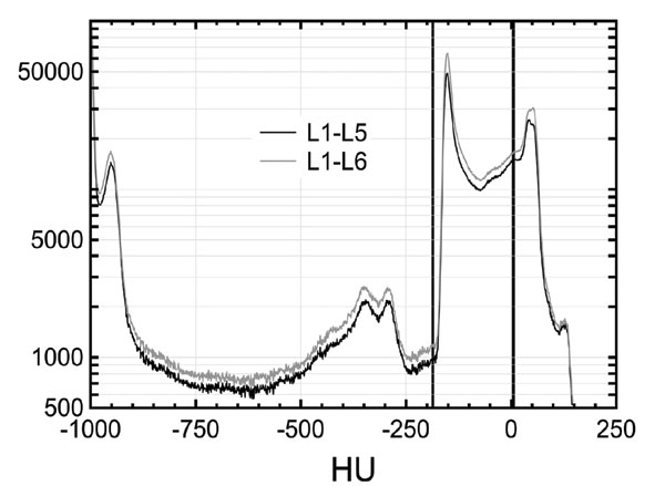
Background: Obesity studies involving animal models require a method for adipose tissue (AT) amount assessment. This paper focuses on the application of clinical computed tomography (CT) for abdominal obesity assessment in rats as an alternative to dedicated microtomographic systems. Additionally, the authors propose L1-L6 instead of L1-L5 region of interest (ROI) usually used for the intra-abdominal adipose tissue (IAAT) assessment and they check if the applied X-ray energy influences results.
Methods: 16 Wistar rats with different body mass (BM) were involved in the study. The animals were scanned by CT to achieve three-dimensional images which were subsequently analyzed for AT amount. AT was identified on the basis of fixed Hounsfield unit scale. Two X-ray tube voltages were tested: 80 kVp and 120 kVp. The results were compared to the fat pads mass (FPM) extracted after animal sacrifice. FPM was also correlated to BM.
Result: The correlation between FPM and BM was statistically non-significant (r=0.3131, p=0.2376). AT amounts obtained for different CT X-ray tube voltages (80 kVp vs. 120 kVp) were practically the same (r=0.9996, p<0.001). There was significant correlation between FPM and AT mass based on CT images, regardless of the ROI choice. Correlation coefficients amount to r=0.932, p<0.001 and 0.945, p<0.001 for L1-L6 and L1-L5, respectively.
Conclusion: BM is not a good descriptor of abdominal obesity. The X-ray beam energy and the choice of ROI do not influence the results considerably. CT allows for fast and reliable IAAT amount assessment in rats.
Keywords: Obesity in rats, fat quantification, computed tomography, image histogram, X-ray, beam energy, body mass.
Graphical Abstract
Call for Papers in Thematic Issues
Intelligent Information Retrieval for Multispectral and Hyperspectral Imaging
Multispectral and hyperspectral imaging is an interdisciplinary research area, widely focuses on spectral, spatial, and temporal data. It offers a plethora of opportunities to efficiently analyze the vast area of the earth's surface. However, there are numerous factors such as dimensionality and size of the hyperspectral data, lesser training samples, ...read more
Machine and Deep Learning in Medical Image Diagnosis
Medical Image Processing regards a set of methodologies that have been developed over recent years with the purpose of improving medical image quality, improving medical data visualization, understanding, and assisting medical diagnosis, and so on. The application of machine and deep learning methods in sensing and imaging can potentially have ...read more
Quantum Machine Learning for Medical Data and Imaging
Quantum computing has promised a significant speedup in certain computationally intensive tasks that are intractable on classical computers. Researchers in quantum machine learning, such as those working in computer vision, image processing, biomedical analysis, and related topics, may play an important role in comprehending and working on complicated medical data, ...read more


 12
12 3
3 1
1